Wild bird migration and the movement of Avian Influenza Virus
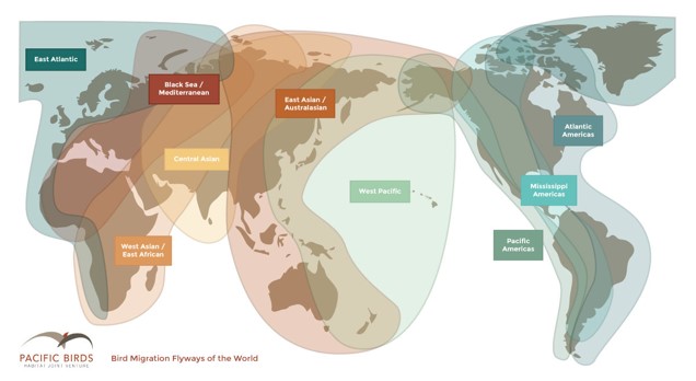
Emily Martin, Avian Pathologist, Animal Health Laboratory, University of GuelphJane Parmley, Associate Professor, Department of Population Medicine, University of Guelph Avian Influenza (AI) is caused by type A influenza virus (family Orthomyxiviridae, genus Influenzavirus A) classified into subtypes by 2 surface proteins: hemagglutinin (H1-H16) and neuraminidase (N1-N9). As an RNA virus, errors are made during replication that cause the virus to change and evolve. Sometimes these errors are minor but other times larger pieces of genetic material can be traded with another AIV virus, increasing the risk of the virus becoming highly pathogenic (causing severe disease in poultry) or able to infect mammals (i.e. swine, humans). Avian Influenza Viruses (AIV) can be grouped as low-pathogenic (LPAIV) or highly-pathogenic (HPAIV, H5 and H7 subtypes). In countries such as Canada where HPAIVs are not endemic and are considered a Foreign Animal Disease (FAD), flocks are destroyed when HPAIV is identified to prevent further spread of the virus. The low pathogenic subtypes of H5 and H7 are handled in the same manner as HPAIV as these subtypes can transform into HPAI as the disease spreads through the flock(s). In order to manage the risk of poultry flocks, or swine and humans, contracting this virus, potential risk factors must be considered. One of these risk factors includes wild birds. AIV naturally infects wild birds and most wild bird species do not show symptoms of infection. All known subtypes of AIV have been isolated from wild birds and LPAIVs are most common. The virus is carried and replicates in the intestinal and respiratory tracts, spreading via feces, saliva, and nasal secretions. Domestic poultry are highly susceptible to AIVs. If flocks are infected with LPAIV the birds may have mild symptoms but the virus is more likely to transform to HPAIV. If a flock is infected with a HPAIV then the birds tend to have severe symptoms and high mortality. This has significant impacts including economic loss, international trade sanctions, and potential spread to humans and other livestock. Since wild birds can carry AIV without symptoms, they can potentially pass AIV to other wild birds, including during migration. There are 8 migratory pathways worldwide and many of these overlap (Figure 1). This creates the potential for AIV to emerge and evolve as well as to move long distances when spread to other migratory or non-migratory birds, including poultry. Previous research tracking H5N1 HPAI between migratory bird flyways identified a correlation between wild bird migration and H5N1 HPAI infections in people. This suggests that transmission is strongly associated with avian migration. Most wild bird samples are collected during the fall and spring migrations with identification of AIVs being more likely during the fall migration and at higher latitudes. It is thought this pattern of identification occurs due to the larger number of naïve, juvenile birds in the population during the fall migration. As the juveniles fly south, they become infected, recover, and are less likely to be carrying the virus once they reach lower latitudes. Wild birds are tested annually in Canada.
Figure 1. The 8 migration pathways. (https://pacificbirds.org/birds-migration/the-flyways/)
There are certain migratory birds, predominantly ducks, followed by geese and gulls that are mainly responsible for annual movement and evolution of AIV. The virus is stable and can survive in the environment for weeks to months. One study showed that AIV in duck feces remained infective for at least 30 days at 4 °C and for 7 days at 20 °C. The aquatic environment is also a potential source of infection for other birds or mammals. While research has provided a better understanding of how AIVs move over long distances (i.e. migration pathways) it is still not well understood how AIVs move from wild birds into poultry populations (i.e. closed poultry barns). To track the movement of AIVs, the strains are compared by looking at their genetic structure in comparison with other isolates. There is a world influenza database (GISAID – Global Initiative on Sharing Avian Influenza Data) where the genetic sequences of AIVs can be uploaded and compared to other AIVs. This can be used to compare AIVs from the same and different geographic areas, look for mutations, and determine if there is movement into bird or mammalian populations (i.e. swine or humans). Part of this comparison consists of constructing a phylogenetic (family) tree of the viruses to see how they cluster into groups; viruses that are more closely related will cluster closer together. As more information is gathered, we can have a clearer picture of how AIVs move within and between species of birds and mammals and how the viruses change over time. It should be noted that when humans become infected with Influenza A (the same group of viruses as AIV), virus mutations or recombinations can occur and birds or swine can then be infected with the virus from humans (and vice versa). That is why poultry workers are encouraged to have a ‘flu shot’ every year to protect themselves and their flocks. Please be aware that there is an ongoing HPAI epidemic in Europe. For more information, please refer to the following links: https://www.efsa.europa.eu/en/efsajournal/pub/9979https://www.thepoultrysite.com/subject/health-disease/diseases/avian-influenza-bird-fluhttps://wahis.oie.int/#/events
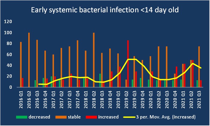
Early systemic bacterial infections (<14 days) were generally stable this quarter as reported by practitioners. Escherichia coli was identified by practitioners. The AHL reported confirmed cases due to E. coli with or without E. cecorum, P. aeruginosa or C. perfringens.Other causes of early mortality (<14 days) was reported as stable to decreased. Late systemic bacterial infections (>14 days) were reported as overall stable with some practitioners seeing a decrease and one reporting an increase in cases. E. coli, Enterococcus cecorum and Clostridium sp were the main bacteria isolated. AHL reported increased numbers of cases attributed to E.coli alone or in various combinations with E. cecorum, S. aureus, S. typhimurium or C. perfringens. AHL reported a decrease in bacterial osteomyelitis and tenosynovitis this quarter that were namely due to E.coli and S. aureus or diagnosed on histopathology. One practitioner noted an increase in Salmonella sp isolation. Isolates listed included group B, D, and S. enteritidis (US source). Reovirus-associated lameness cases were decreased to stable by practitioners. AHL reported an decrease in confirmed cases. A number of different genotypes were detected. In birds <14 days, variants identified included H, K and SK_R38. In birds >14 days, variants included F, A, SK_R38 and Ontario classic 10-076656. Escherichia coli and E. cecorum alone or in combination were identified as the main causes of bacterial lameness this quarter with most practitioners reporting stable to increased prevalence.
The bars represent the proportion (%) of veterinarians who report the number of cases seen in a quarter as decreased, stable or increased compared to historical expected numbers of cases.
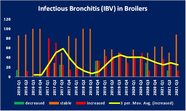
The number of cases of nutritional and developmental lameness was reported as stable to increased by the majority of responders. Rickets, general micronutrient deficiency (riboflavin deficiency, phosphorus deficiency and calcium deficiency) being noted as diagnoses by practitioners. AHL also reported cases of tibial dyschondroplasia and tibiotarsal rotation. Ascites was reported as stable to increased by practitioners this quarter. One practitioner suggested that ascites may be increased due to an association with some types of bronchitis virus like DMV. Coccidiosis and necrotic enteritis cases were reported as stable to increased by the majority of practising veterinarians. One practitioner noted a decrease in coccidiosis cases. The AHL also reported decreased coccidiosis cases, identifying the lesions in the small intestine, ceca or both. Necrotic enteritis esions were only reported in birds <14 days. Inclusion body hepatitis (IBH) cases were reported to be stable to increased by the majority of respondents and AHL this quarter. Genotyping on confirmed cases identified mainly FAdV8b of the species E viruses in birds <14 days. Different combinations of isolates of FAdVAC, FadVE and FadVD in this age group. For birds >14 days, AHL reported FAdVE, FadVD and FAdVAC/FAdVE in confirmed cases or some combination of the 3 species. The number of flocks with infectious bronchitis virus (IBV) were reported as stable to increased this quarter by the majority of poultry practitioners and AHL. The DMV strain and the Mass and Conn vaccine strains were identified. Infectious bursal disease virus (IBDV) infections were reported as stable to increased this quarter by practicing veterinarians and AHL although there is a increase in testing (3 vaccine and 1 USA PA105-2014 field strain).
The bars represent the proportion (%) of veterinarians who report the number of cases seen in a quarter as decreased, stable or increased compared to historical expected numbers of cases.
Submissions to the AHL of confirmed runting and stunting syndrome caused by astrovirus were slightly decreased this quarter. Proventricular and ventricular lesions were reported as stable by AHL. One practitioner reported an increase in proventricular dilation in a flock of guinea fowl fed excessive grit. One practitioner reported a case of salt toxicosis in 3 week old birds. Condemnation issues were reported as stable by veterinarians this quarter and included cellulitis, woody breast, ascites, air sacculitis, hepatitis and ascites.
Broiler-Breeders
Early systemic bacterial infections (<14 days) with and without associated yolk sacculitis were reported as stable to increased by practitioners with one noting a decrease in cases. E. coli continues to be the most common isolate and was reported combined with C. perfringens by one practitioner with high mortality (enteritis and liver lesions). AHL reported E. coli with or without E. cecorum. Pre-lay mortality (> 14 days, < 20 weeks) at AHL identified E. coli alone or in combination with E. cecorum. One submission included E. coli, E. cecorum and S. aureus. Other causes of early mortality reported by practitioners and AHL were stable this quarter and included dehydration, starveout, mycotic pneumonia and intussusception (with coccidiosis). In-lay bacterial septicemia was reported as stable to increased by practitioners, mainly due to E. coli, G. anatis and E. cecorum. G. anatis is being seen more frequently as a mixed infection. Similarly, AHL reported E.coli, S. aureus, E. cecorum and G. anatis in various combinations. One practitioner noted a rare vaccine strain of fowl cholera was identified. Lameness was reported as stable by the majority of practitioners this quarter with one report of an increase in lameness due to nutritional causes. S. aureus was the most common bacterial cause of lameness reported by practitioners and AHL (tenosynovitis) along with some mixed infections with E. coli and E. cecorum. Occasionally, Pasteurella sp (usually vaccine strain) was isolated. One practitioner noted an increase in developmental lameness with about 1-2% of the flock developing fractures. It was determined there was growth plate damage, cause undetermined) with a subsequent effect on bone strength leading to fractures. IBV infections were reported as stable by the practitioners and at AHL. One practitioner reported two cases of cystic oviducts which presented as chronic mortality in the summer heat. One practitioner commented that a sudden spike in mortality due to IBV is often seen with the 2nd infection and associated with the Delmarva or CU strains. AHL also reported an increase in cystic oviducts. The number of coccidiosis and necrotic enteritis cases reported by practitioners was stable this quarter. One practitioner commented that coccidiosis is frequently seen in flocks with high bacterial challenges. AHL reported a slight increase in both coccidiosis including E. necatrix cases, affecting the small intestine and ceca. An increase in E. necatrix may be due to a change in vaccine. White chick syndrome (WCS) was reported by AHL (2 cases). Poultry practitioners reported a variety of Salmonella isolates including S. Livingstone, S. Infantis, S. Hadar, S. Putten and S. Mbdanka. One practitioner noted they did not isolate any Salmonella sp this quarter.
Layers
Bacterial peritonitis/salpingitis due to E. coli, G. anatis and E. cecorum was stable. AHL reported a few of cases of septicemia, and yolk sacculitis (diagnosed on histology). Early systemic bacterial infections (<14 days) were stable. Cases of pre-lay mortality (>14 days, < 20 weeks) at AHL identified E. coli. Focal duodenal necrosis (FDN) was reported as stable to increased by practitioners. AHL reported one case diagnosed by histology. IBV was reported as stable to increased by practitioners and AHL who reported one case of cystic oviducts. Additional samples submitted to AHL Virology identified CU82792, IBV_Mass-MA5 vaccine, IBV_Conn vaccine and IBV_DMV_ON_5—077145. Necrotic enteritis and coccidiosis cases were reported as stable to increased by practitioners and AHL. E. necatrix was identified in one case by AHL. Other diagnoses made by AHL included; salpingitis, cannibalism, gangrenous dermatitis (E. coli, S. aureus), fatty liver and vent trauma.
Turkeys
Early systemic bacterial infection <14d were stable to increased. E.coli and Salmonella sp. were reported by practitioners. E. coli in combination with E. cecorum, P. aeruginosa, S. Hadar and S. Uganda were noted by AHL. Other causes of early mortality < 14d were stable to decreased this quarter. Excessive beak trimming was listed as a cause. Late systemic bacterial infection >14d were stable to increased with E. coli and Salmonella Agona being reported by practitioners. AHL reported isolation of E.coli or E.coli with other bacteria (S. aureus, E. cecorum, C. perfringens). Two practitioners reported an increase in mycotic respiratory disease and AHL reported an increase in mycotic pneumonia. ORT was reported as stable to decreased by practitioners. One practitioner noted a case of fowl cholera in their practice. Necrotic enteritis and coccidiosis were reported as stable to increased by the majority of practitioners. Enteritis was reported as stable to increased this quarter by practitioners. AHL reported one case due to E. coli and S. Uganda. AHL reported some cases of tibial dyschondroplasia, tenosynovitis (due to E. coli or on histology), valgus/metatarsal bone rotation, ionophore toxicity (suspicious), mycotic osteomyelitis, dilated cardiomyopathy and lesions suspicious for turkey viral hepatitis. Veterinarians and AHL reported tenosynovitis due to reovirus was stable to increased this quarter. One producer culled almost half of their flock of 16 week old toms. One case at AHL was positive for the PA_13-22342 field strain and some were positive on histology (lymphoplasmacytic tenosynovitis and/or epicarditis). Aggression and cannibalism was reported as stable to increased by practitioners. Histomoniasis (blackhead) was reported as stable to increased. One practitioner reported a couple of severe cases. Biosecurity is important to avoid moving the parasite between farms. One practitioner reported a case of suspected mycotoxicosis based on histology. Poultry practitioners reported a variety of Salmonella isolates including S. Liverpool, S. Uganda, S. Schwarzengrund, S. Senftenberg, and S. Agona.
Rural/Backyard/Non-Quota Flocks
AHL continues to receive increasing submissions from backyard flocks. The number of backyard/urban poultry has increased due to COVID-19. A significant increase in intestinal parasitism was reported by AHL with primarily coccidiosis (small intestine and ceca) including E. necatrix reported. Nematodes, tapeworms and cecal tetratrichomonas were also identified. Marek’s disease continues to be diagnosed. One case of ILT was diagnosed by AHL; a Niagara-like field strain, CAGG cluster. The virology lab had 3 PCR positive cases; 2 USA vaccine-like, 1 TCAA cluster. AHL reported a large variety of diagnoses: necrotic enteritis, adenocarcinoma, reproductive issues (salpingitis), bacterial septicemia, urate nephrosis, right heart failure, vertebral osteomyelitis, emaciation, amyloidosis and uveitis. An increase in water fowl cases were reported by AHL with a variety of diagnoses: lung/liver hemorrhage, gizzard foreign body, fatty liver, parasitism (peritonitis w. trematode), septicemia and renal tubular necrosis.
Thank You! We thank the following poultry veterinarians who completed the veterinary survey: Dr. Elizabeth Black, Dr. Peter Gazdzinski, Dr. Shahbaz Haq, Dr. Geneviève Huard, Dr. Anastasia Novy, Dr. Mike Petrik, Dr. Joanne Rafuse, Dr. Kathleen Sary, Dr. Ben Schlegel, Dr. Chanelle Taylor, Dr. Lloyd Weber, Dr. Alex Weisz, and Dr. Jessalyn Walkey.
 Avian Influenza Viruses (AIV) can be grouped as low-pathogenic (LPAIV) or highly-pathogenic (HPAIV, H5 and H7 subtypes). In countries such as Canada where HPAIVs are not endemic and are considered a Foreign Animal Disease (FAD), flocks are destroyed when HPAIV is identified to prevent further spread of the virus. The low pathogenic subtypes of H5 and H7 are handled in the same manner as HPAIV as these subtypes can transform into HPAI as the disease spreads through the flock(s). In order to manage the risk of poultry flocks, or swine and humans, contracting this virus, potential risk factors must be considered. One of these risk factors includes wild birds. AIV naturally infects wild birds and most wild bird species do not show symptoms of infection. All known subtypes of AIV have been isolated from wild birds and LPAIVs are most common. The virus is carried and replicates in the intestinal and respiratory tracts, spreading via feces, saliva, and nasal secretions. Domestic poultry are highly susceptible to AIVs. If flocks are infected with LPAIV the birds may have mild symptoms but the virus is more likely to transform to HPAIV. If a flock is infected with a HPAIV then the birds tend to have severe symptoms and high mortality. This has significant impacts including economic loss, international trade sanctions, and potential spread to humans and other livestock. Since wild birds can carry AIV without symptoms, they can potentially pass AIV to other wild birds, including during migration. There are 8 migratory pathways worldwide and many of these overlap (Figure 1). This creates the potential for AIV to emerge and evolve as well as to move long distances when spread to other migratory or non-migratory birds, including poultry. Previous research tracking H5N1 HPAI between migratory bird flyways identified a correlation between wild bird migration and H5N1 HPAI infections in people. This suggests that transmission is strongly associated with avian migration. Most wild bird samples are collected during the fall and spring migrations with identification of AIVs being more likely during the fall migration and at higher latitudes. It is thought this pattern of identification occurs due to the larger number of naïve, juvenile birds in the population during the fall migration. As the juveniles fly south, they become infected, recover, and are less likely to be carrying the virus once they reach lower latitudes. Wild birds are tested annually in Canada.