
Ontario Animal Health Network (OAHN) Bovine Expert Network Quarterly Veterinary Report
Global Surveillance Updates: New World screwworm in Texas
After many months of anticipating its introduction, New World Screwworm (NWS) was confirmed in a calf in the United States in early June 2026. Since then, multiple domestically acquired cases have been confirmed in livestock across South Texas counties, and one detection in New Mexico. This follows the re-emergence of NWS across Central America and Mexico since 2023.
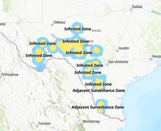

Important Info for Ontario Veterinarians:
- NWS is not present in Canada
- While the cold Canadian climate prevents NWS from permanently establishing, detections in Texas have increased concern about introduction through animal movements
- CFIA has banned the import of livestock, including horses, cattle, bison, sheep, goats and swine, that originate from or have travelled through Texas within the prior 21 days.
- Know the signs of screwworm, which may include:
- wounds that enlarge or fail to heal
- discharge or foul-smelling odour from a wound
- visible maggots in a wound or body opening
- pain, discomfort or irritation
- decreased appetite, reduced milk production or animals separating from the herd
- If suspected, contact your local district CFIA office for guidance
- Any suspect larvae should be submerged into 70% ethanol to preserve them
Q1 Bovine Data from the Animal Health Laboratory
Bovine Viral Diarrhea Virus – Associated Abortions
Case 1: PCR-Confirmed BVDV Abortion in a Holstein Fetus with Minimal Pathologic Lesions
History: Holstein fetus. Abortion 6 months pregnant. Multiple abortions in last 1-2 months.
Pathology: Minimal gross and histologic lesions.
Ancillary testing: BVDV PCR positive (Ct 27), no significant agents on culture and BHV-1, Leptospira, and Neospora negative.
Summary: Despite the lack of histologic lesions, the detection of BVDV in fetal tissues is considered significant and this was diagnosed as abortion due to BVDV.
Case 2: BVDV-Associated Fetal Loss in a Holstein Herd with Clustered Abortions and Premature Calving Events
History: Holstein fetus. 3 abortions in the last week. 6 cows/heifers have also calved 3 weeks early within the last few weeks with live calves.
Pathology: Minimal gross lesions, histology detected thymic cortical atrophy and lymphocytic myocarditis with fibrosis.
Ancillary testing: BVDV PCR positive (Ct 30), 2+ Bacillus licheniformis from the amnion.
Summary: This case had detection of BVDV by PCR as well as corroborating histologic lesions. In discussion with the submitting clinician, it was confirmed this herd is not vaccinated for BVDV. Secondary bacterial infections can occur in cases of BVDV abortion.
Read the comprehensive case report at: Bovine abortion due to BVDV infection | Animal Health Laboratory
BVD Diagnosis: A Quick Review
1. PCR (for Virus Detection)
Sample types:
– EDTA blood, serum, plasma
– Tissues, ear notches
– Milk (unfrozen, including bulk tank)
Primary test for detecting persistent (PI) and acute infection
Pooling (typically 5-10 per pool) possible when used for screening
2. Virus neutralization (VN) test (for Antibody Detection)
Sample types: serum
Can be used to demonstrate evidence for:
- Previous or recent exposure to BVDV type 1 or type 2 (single sera test)
- Seroconversion (paired sera collected 2-4 weeks apart)
- In utero infection (sera collected at birth prior to colostrum ingestion)
*Vaccine status and product important for interpretation
3. Immunohistochemistry
Sample type: formalin-fixed tissue when fresh tissue not available
Used for individual diagnostics at postmortem
For more information:
LabNote 1- Summary of bovine viral diarrhea virus (BVDV) testing at the AHL | Animal Health Laboratory
Other Cases of Interest
Bilateral Functional Pheochryomocytomas in a Scottish Highland Cow
History: 17-year-old Scottish Highland cow. Presented on the morning of April 20th with apparent interest in eating grain and drinking but did neither- went to food and water but did not prehend. She walked compulsively around the pen with no evidence of ataxia or weakness. No excessive salivation, tongue tone good, tail reflex strong. Increased respiratory rate and increased rectal temperature. Mildly increased breath sounds on auscultation. Next morning found in sternal recumbency and depressed. She was able to get up but appeared weak but not ataxic. No interest in food or water and was euthanized.
Pathology: An 11 cm by 9 cm by 5 cm mass expanding one pole of the left adrenal gland. Center of the right adrenal gland is expanded by a soft, red, 2 cm diameter nodule. Neuroendocrine neoplasm confirmed bilaterally on histology.
Ancillary testing: Rabies testing performed-negative. Synaptophysin IHC confirmed both masses were pheochromocytomas.
Summary: Pheochromocytomas can be functional, and if they are, they produce catecholamines. This can lead to clinical signs including listlessness, anorexia, weakness, tachypnea, and pyrexia. No other cause for the clinical signs was noted, therefore a functional pheochromocytoma is the presumed cause of the clinical presentation.
Pulmonary carcinoma in an Adult Beef Cow
History: Adult beef cow. Pneumonia symptoms, frothing at the mouth.
Pathology: Extensively effacing the lung as well as the pleura there is an epithelial neoplasm. There is evidence of vascular invasion and tumor emboli in multiple organs including the heart.
Ancillary testing: None
Summary: The significant effacement of the lung by a carcinoma is the presumed cause of the respiratory clinical signs. Given the tissues examined and clinical signs, the lung is the presumed primary site of development of the neoplasm, but other tissues not available for examination such as the uterus cannot be ruled out.
Stats for Q1
There were a total of 1853 bovine submissions in Q1 2026. Of these, a total of 210 submissions to the AHL had a pathology component:
- 70 postmortem cases and 140 send-in cases (including 46 meat inspection)
- 85 dairy, 73 beef, 6 not-specified
- Animals with the commodity not specified were down significantly from Q4 2025 (25).
- 132 submissions had a definitive or presumptive diagnosis, 32 did not have a specific diagnosis (10 abortion work-ups, 5 young calves, 14 older calves, 3 adults)
- The vast majority of these cases were send-in. For most submissions, a very limited set of tissues had been sampled for histopathology or for culture, and history was often minimal.
- Insufficient clinical history (none) was associated with 4 submissions.
| Category | Number Submissions |
| Young calves (under 2
months) |
56 |
| Older calves (2 months-2
years) |
55 |
| Adult cattle (> 2 years) | 26 |
| Reproductive Loss | 27 |
| Meat inspection | 46 |
Salmonella Report
In total, 262 bovine submissions had bacterial culture performed (non-milk), generating 388 cultures. Salmonella spp. were isolated from 7 submissions, representing an estimated 6 premises.
Salmonella Dublin was isolated from 5 submissions, representing an estimated 4 premises (first 2 cases listed are from the same premise). Three of these cases, representing 2 premises, had previous positive submissions.
- Dairy calf, poor doing since birth then developed icterus and sepsis. Lung PCR positive and liver and kidney S. Dublin positive on culture.
- Dairy calves, chronic pneumonias. Histologic lesions nonspecific but S. Dublin isolated from the lung.
- Dairy calves, scours in all ages. Histologic lesions of sepsis, lung and intestine positive for S. Dublin.
- Dairy calf, peritonitis. Histologic lesions of enteritis with S. Dublin isolated from the intestine.
- Dairy calf, diarrhea and resp signs. No histology, S. Dublin isolated from feces.
There were 4 bovine submissions that had Salmonella Dublin PCR performed, generating 1 positive submission (17-day-old dairy calf with sepsis). This animal is also captured in the culture data above.
Salmonella Anatum (1 submission) detected on fecal culture
Salmonella Agona (1 submission) detected on fecal culture
This summary has been compiled by Dr. Dominique Comeau, Animal Health Laboratory (AHL) from diagnostic submissions to the AHL Guelph and Kemptville locations.
Mycobacterium avium paratuberculosis (MAP) Testing: Laboratory Summaries
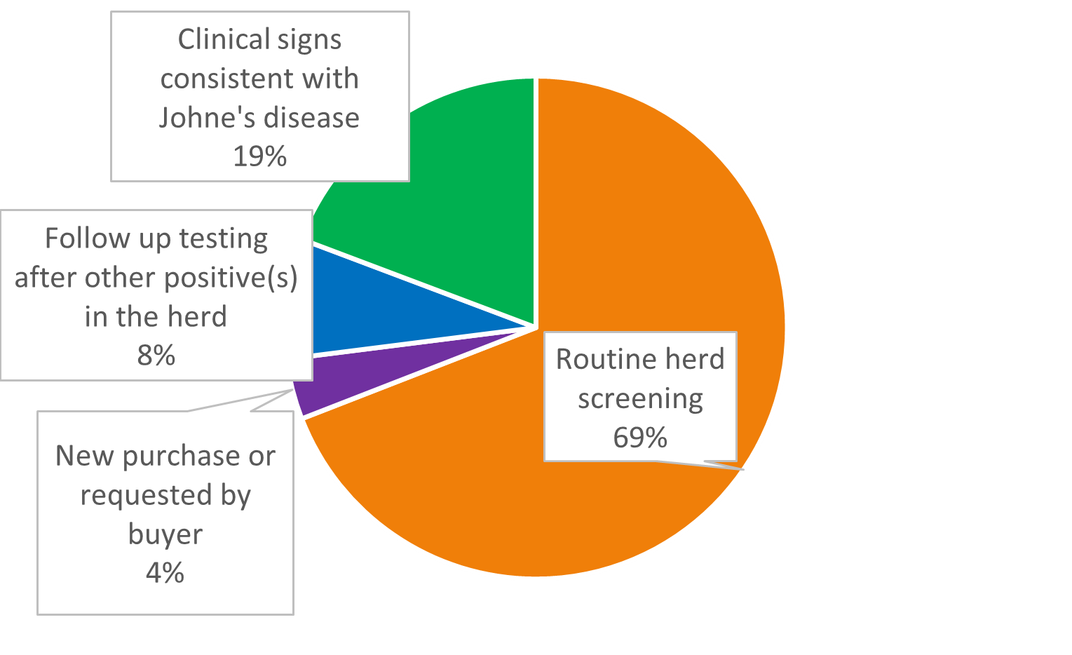
To start Q1, the OAHN bovine network identified a review of Johne’s disease testing conducted in Ontario as a priority area for review. The network last summarized testing in Ontario in 2021. Data from Lactanet and the Animal Health Laboratory was reviewed to look for trends and current testing occurring in dairy and beef cattle. Testing may be conducted for many reasons – routine screening or in response to clinical disease.
Johne’s Testing at the Animal Health Laboratory
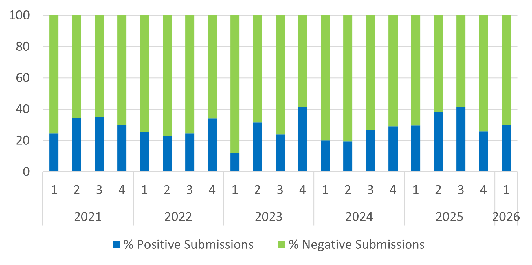
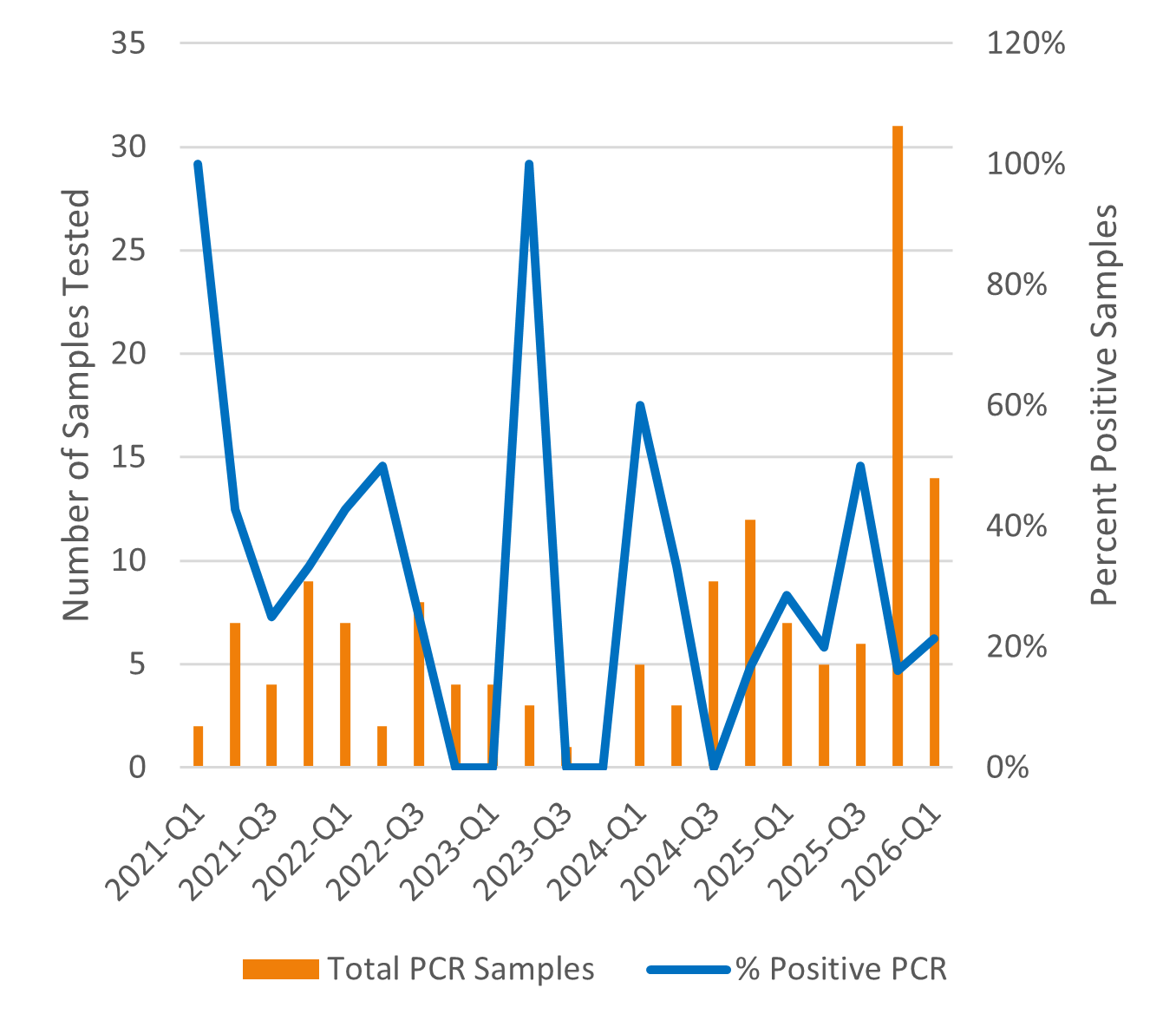
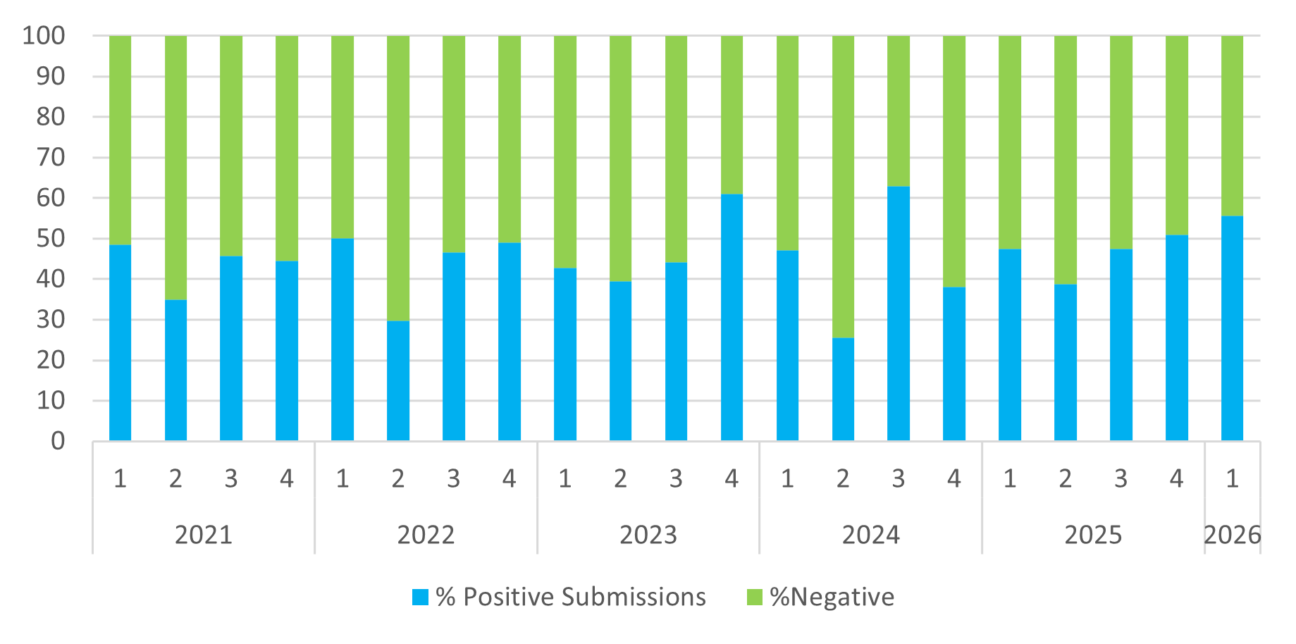
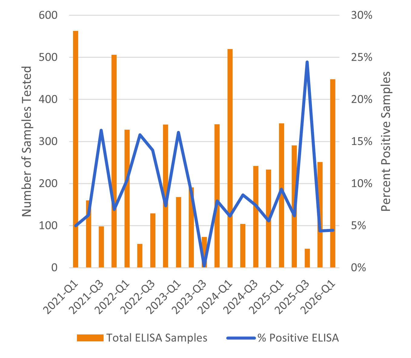
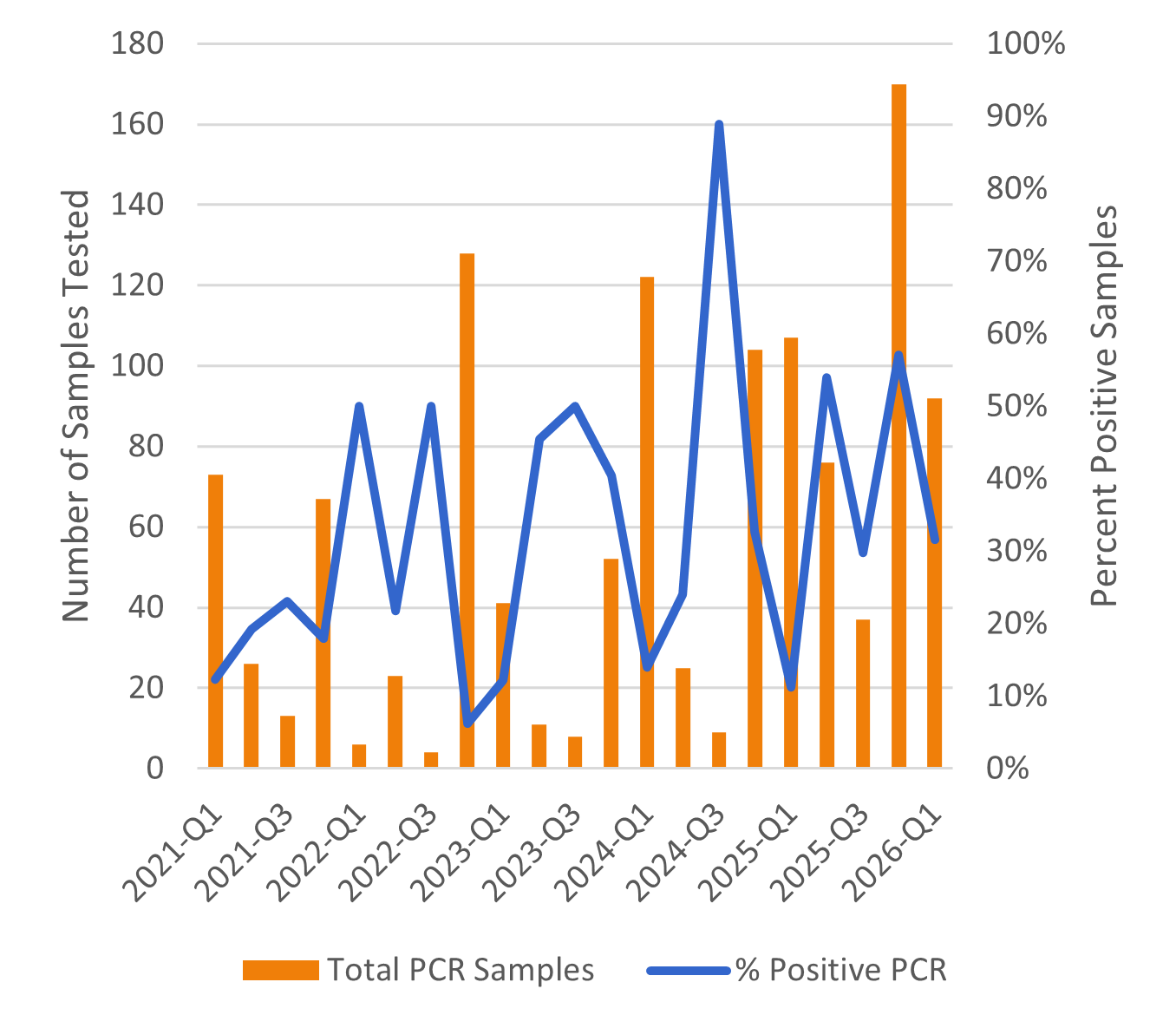
The AHL provides the ELISA test on serum and the PCR test on feces. Between January 1, 2021 and March 31, 2026, a total of 9,296 tests for Mycobacterium avium paratuberculosis were performed.
Dairy Results
|
Number of Tests – DAIRY |
||
| ELISA | PCR | |
| 2021 | 718 | 22 |
| 2022 | 429 | 21 |
| 2023 | 424 | 8 |
| 2024 | 361 | 29 |
| 2025 | 465 | 49 |
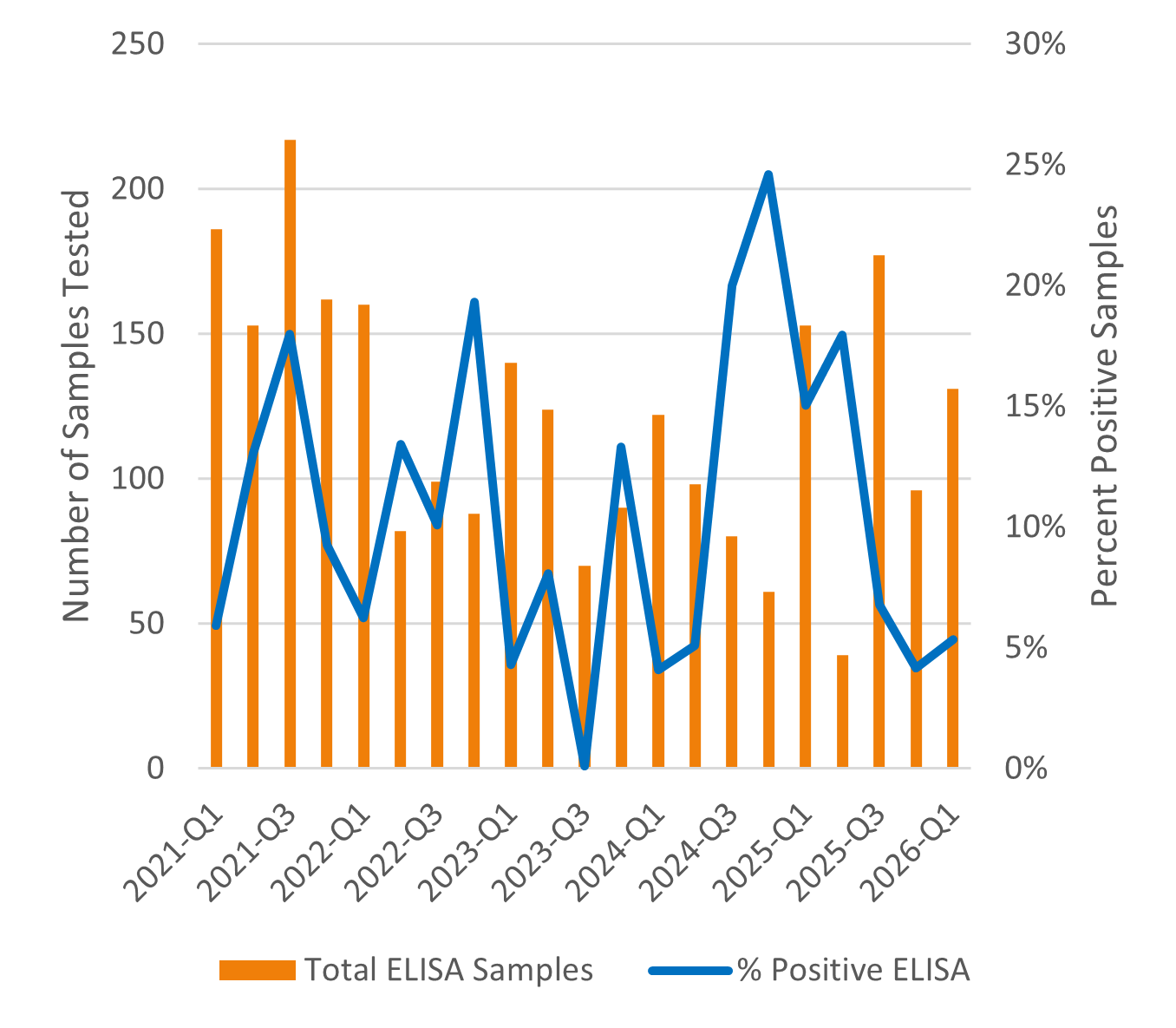
Beef Results
| Number of Tests – BEEF | ||
| ELISA | PCR | |
| 2021 | 1327 | 179 |
| 2022 | 854 | 161 |
| 2023 | 773 | 112 |
| 2024 | 1099 | 260 |
| 2025 | 930 | 390 |
Johne’s Disease Passive Surveillance: What Lactanet Milk ELISA Data Tells Us
Johne’s disease remains widely distributed in dairy herds, yet relatively few producers are actively monitoring it. Lactanet milk ELISA data suggest that infection is commonly present at the herd level, even when only a small proportion of cows test positive.
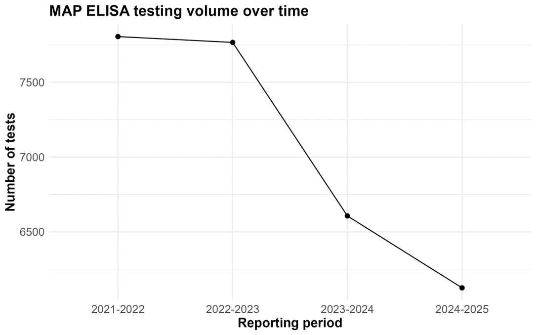
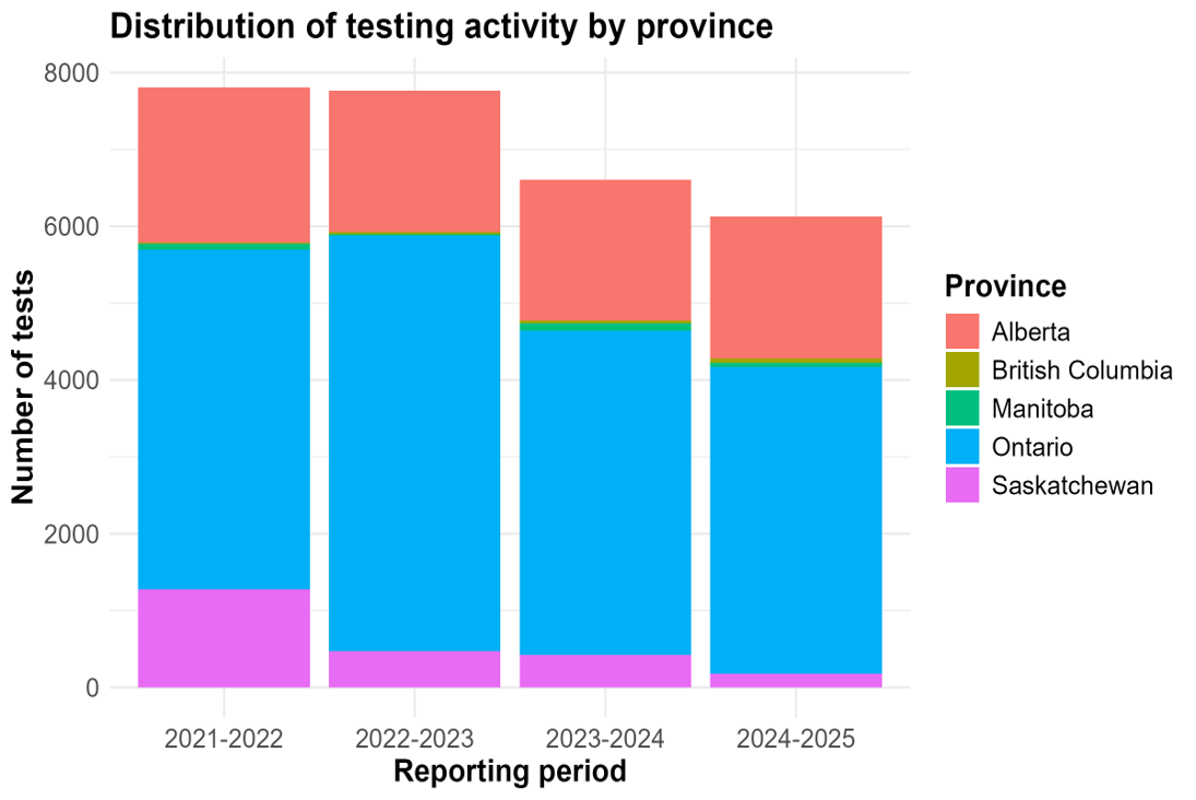
Johne’s disease, caused by Mycobacterium avium subspecies paratuberculosis (MAP), is a chronic infectious disease that can result in reduced production, premature culling, weight loss, and eventual death. Although clinical Johne’s disease represents the visible “tip of the iceberg,” infected herds often contain many more subclinical animals that contribute to ongoing transmission. Between October 2021 and September 2025, Lactanet processed 28,306 milk ELISA tests from 18,035 cows in 240 dairy herds across Ontario and Western Canada. Ontario accounted for the majority of testing activity (57-70% yearly of all submitted samples.
Despite the widespread availability of milk ELISA testing, only a small proportion of dairy herds in these provinces participated in Johne’s surveillance during the study period.
What the Data Show
Cow-Level Results
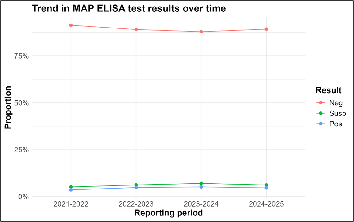
- Overall, 4.5% of tested cows were ELISA positive, while 6.1% were classified as suspect. Individual-animal positivity remained relatively stable throughout the four-year period.
- As expected, positive results were more common in older cows (6.0% positive in 3rd lactation and greater cows versus 3.4% in 1st and 2nd lactation cows). This finding is consistent with the epidemiology of Johne’s disease; cattle are typically infected early in life, but antibody responses may not become detectable for years. Because ELISA sensitivity increases with disease progression, older animals are more likely to test positive than younger infected animals (Nielsen & Toft, 2008).
- Breed-level patterns were also consistent with some field observations. Holsteins showed a positivity rate of 3.5%, while Jerseys demonstrated a higher apparent positivity rate of 6.7%. However, breed comparisons should be interpreted cautiously, as management practices, herd demographics, and testing strategies may also influence observed differences.
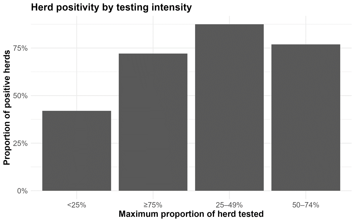
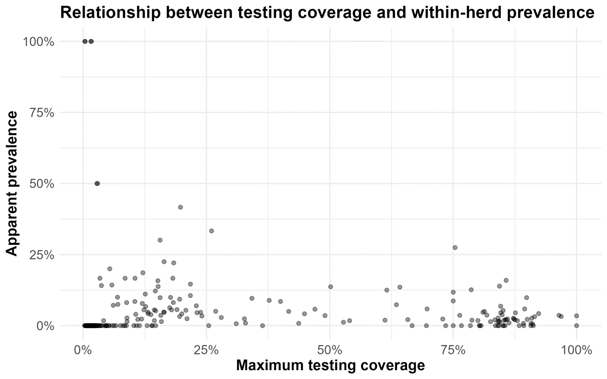
Herd-Level Results While cow-level prevalence appeared relatively low, the herd-level picture was very different. More than half of participating herds (54.6%) had at least one positive animal detected during the study period. Among herds with higher testing coverage (≥50% of cows tested), herd-level positivity increased to 73.0%. This difference highlights an important surveillance principle: the more animals tested, the more likely infection will be detected.
- The majority of herds (62.5%) tested fewer than 25% of their animals, suggesting that herd infection status may be underestimated in many herds due to low testing coverage.
- Despite widespread detection, the burden of infection within most herds remained relatively low. Among well-sampled herds, median within-herd apparent prevalence was only 2.1%, and most herds had fewer than 10% positive animals.
Why It Matters
- Johne’s disease continues to represent a significant animal health and economic challenge for the dairy industry.
- Previous Canadian research estimated herd-level prevalence to exceed 40% of dairy herds (Corbett et al., 2018), while economic losses associated with Johne’s disease were estimated at approximately $35-57 USD per cow annually, equivalent to roughly 0.6-1.0% of gross milk revenuein affected herds (Rasmussen et al., 2021).
- Beyond direct production losses, MAP transmission is closely associated with management practices that also influence other diseases affecting youngstock, including neonatal diarrhea and Salmonella infections. As a result, efforts to control Johne’s disease often support broader herd health objectives.
Why Encourage Milk ELISA Testing?
One of the greatest challenges of Johne’s disease is the prolonged interval between infection and clinical disease. Most infected animals remain undetected for years, and clinical cases often appear only after significant MAP transmission has already occurred within the herd. Milk ELISA testing offers veterinarians and producers a practical surveillance tool that is:
- Convenient, non-invasive, and cost-effective
- Easily incorporated into routine Lactanet milk recording
- Useful for identifying higher-risk animals
- Valuable for tracking herd infection status over time
- Repeated testing improves confidence in identifying infected animals and monitoring trends within the herd.
Key Message
- The Lactanet data reinforce a familiar but important reality: Johne’s disease is commonly detected at the herd level, even though only a small proportion of cows may test positive. The disease is often a “silent robber” of productivity, with many infected animals remaining subclinical for years. The relatively low within-herd prevalence identified in this analysis suggests that many herds may still have an opportunity to limit transmission before infection becomes more widespread.
- Only 240 herds contributed milk ELISA data over the four-year study period, representing a small fraction of the more than 4,400 dairy herds in Ontario and Western Canada (Government of Canada, 2025). Increasing surveillance participation represents an important opportunity for the dairy industry.
- Veterinarians play a key role in helping producers establish herd status, develop appropriate testing strategies, and implement practical Johne’s disease control measures. As with any herd disease, effective control starts with measurement: if we do not test for Johne’s disease, we cannot effectively manage it.
This summary has been compiled by Dr. Elouise Molgat, Lactanet from milk testing completed from herds in Ontario, Manitoba, Saskatchewan, Alberta, and British Columbia.
Practical resources for improving calf health
- This tool was designed using systematic review, expert consensus and industry data and covers environment, perinatal management, nutritional management, biosecurity and vaccination. It can be used to facilitate productive discussions about calf health and prevention strategies between producers and veterinarians.
- Check out the supplementary material if you want to try out the questionnaire.
A Framework for Comprehensive Dairy Calf Health Investigations
- This review provides a practical approach to investigating calf health issues, helping veterinarians and producers identify risk factors and improve calf health on farm.