
Ontario Animal Health Network (OAHN) Bovine Expert Network Quarterly Veterinary Report
Global Surveillance Update: European Shamonda Virus
![Figure 1: Map of countries known to be affected with European Shamonda Virus, including Switzerland, Germany, France, Austria, the Netherlands, Belgium, Liechtenstein, Spain, Italy, Luxembourg, Jersey in the British Isles, and Ireland as of October 6, 2026. [Produced using Microsoft CoPilot]](https://www.oahn.ca/wp-content/uploads/2026/10/Euro-Shamonda-Virus.png)
Emergence and Spread in Europe
In mid-June, farmers reported an acute disease in dairy cattle characterized by high herd morbidity, decreased milk yield, diarrhea, fever, and lethargy. After other major viral diseases were excluded as the cause, metagenomic sequencing was used to identify a novel Shamonda virus. Shamonda virus belongs to the genus Orthobunyavirus and the Simbu serogroup. Shamonda virus was first isolated in Nigeria in the 1960s from cattle and Culicoides biting midges. Historically, Shamonda virus has been restricted to Africa, Asia and Australia. Initially referred to as “Shamonda-like virus” in scientific publications, it is now being called European Shamonda virus (SHAV). Two Shamonda virus variants have now been identified in Europe. SHAV-EU1 has been detected in southern Germany, Switzerland, France, and Austria, while SHAV-EU2 has been detected in northern Germany, Belgium, the Netherlands, and northwestern France. Both European variants differ genetically from the original Nigerian Shamonda virus and may represent two separate incursions, although their origin remains unclear and may involve a new introduction, rapid mutation, or reassortment.
One hypothesis is that an infected midge was transported by air travel, with warm summer temperatures in Europe enhancing viral replication and subsequent transmission by European midge species. Major international airports located near intensive livestock production areas may have created conditions that allowed SHAV to emerge and spread.
Clinical Signs and Affected Species
To date, SHAV-EU1 and SHAV-EU2 have caused disease in cattle with high morbidity, reaching up to 100% in some herds, but no mortality has been reported. In one publication from France, some herds experienced up to a 75% reduction in herd milk production. However, reports vary considerably regarding both the magnitude of milk production losses and the proportion of cows with a fever, depression, and diarrhea. Mild, self-limiting respiratory signs have been reported rarely. Clinical signs are typically reported to last between 1-6 days.
Although infections have been reported predominantly in lactating dairy cows, detections have also occurred in horses, sheep, goats, and alpacas. In horses, detections were associated with animals that died after exhibiting signs of central nervous disease. Two alpacas with CNS signs also tested positive in Germany. Sheep and goats were tested as part of surveillance in southern Germany and viral RNA was detected in 7 sheep and 3 goats.
Reproductive Concerns and Future Risks
This is not the first midge transmitted disease to emerge in Europe. In 2011, Schmallenberg virus (SBV), another novel orthobunyavirus named after the town where it was first identified, emerged in Europe. The virus causes congenital malformations and stillbirths in cattle, sheep, and goats. Since 2023, Europe has also experienced the rapid spread of bluetongue virus, particularly serotype 3, through Western Europe and into the United Kingdom. A recent article expressed concern that the concurrent circulation of SHAV-EU1, SHAV-EU2, and SBV could result in the emergence of a reassortant strain with altered host range, transmissibility, or pathogenicity1.
Many uncertainties remain regarding the impact of SHAV. Early in the outbreak, the veterinary community identified concerns for potential fetal effects, including abortion, malformation, or stillbirths, similar to those associated with SBV and Bluetongue virus. Evidence supporting these concerns is beginning to emerge. The virus has been detected in bovine fetuses, indicating it can be transmitted through the placenta. A recent Swiss publication reported 22 aborted calves that have tested positive in the spleen, brain, or both. In addition, a publication from France identified viral RNA in tissues from 18 fetuses, providing evidence of association between infection and reproductive disease in cattle. The long-term impacts on fertility have yet to be quantified. Currently, there are no specific vaccines or treatments available.
Key Points for Veterinarians:
- At this time, there is no evidence of SHAV-EU1 or SHAV-EU2 in North America
- Occurs in cattle and other livestock with high morbidity but low mortality
- Risk for abortion remains to be characterized but has been documented
- Likely being spread by midges
- Concerns about any potential vector-mediated disease should be reported to CFIA. Find your local district office:
- Veterinarians can find more information on SHAV from the Friedrich Loeffler Institut in Germany at Shamonda-Virus (SHAV)
References:
- New midgeborne virus hits Europe. Veterinary Record. 2026. doi:10.1002/vetr.71176
- Wyler M, et al. Identification, Isolation and Genomic Characterization of an Emerging Shamonda Virus Variant Associated with Bovine Disease in Switzerland. Viruses. 2026. doi:10.3390/v18101086
- Kelleci M, et al. Detection of a novel Shamonda Orthobunyavirus in dairy cattle, France, June 2026. Euro Surveill. 2026. doi:10.2807/1560-7917.ES.2026.31.33.2600689.
Q2 Bovine Data from the Animal Health Laboratory
Stats for Q2
Total bovine submissions received: 1,973
Of these, 159 submissions (8.1%) included a pathology component.
Postmortem cases: 55
Send-in cases: 104 (including 34 meat inspection cases)
Pathology Submissions by Category
Young calves (< 2 months): 38
Older calves (2 months to 2 years): 29
Adult cattle (> 2 years): 30
Reproductive loss (abortion): 28
Meat inspection: 34
Production Type
Dairy: 66
Beef: 48
Not specified: 11
Diagnostic Outcomes
Definitive or presumptive diagnosis obtained: 100 submissions
No specific diagnosis: 25 submissions
Reproductive loss: 11
Young calves: 4
Older calves: 4
Adult cattle: 6
Note: Failure to reach a specific diagnosis was most common in reproductive loss investigations. In some cases, limited sample submission (e.g., placenta only, or fetus submitted without placenta) likely contributed to the inability to identify a cause.
Case Highlights
Case 1: Congenital Interstitial Cell Tumor in a Calf
History: 3-week-old Angus/Holstein cross calf. Owner reports it was born with an enlarged testicle. Surgical removal of the testicle was performed. Testicle was cut open and appearance was abnormal. Trauma was ruled out based off appearance. Hyperplasia or neoplasia was suspected based on appearance.
Pathology: Histologic sections of the testis were extensively effaced by a neoplasm composed of polygonal cells forming sheets and cords with minimal stroma. There was minimal pleiomorphism, and no mitotic figures were noted.
Summary: Congenital interstitial cell tumors are extremely rare in cattle, and congenital neoplasms overall are uncommon but reported sporadically. Interstitial cell tumors in general are benign and metastasis is exceedingly rare.
*Read more about this case in the AHL Newsletter
Case 2: Supernumerary Limb in a Heifer Calf
History: 1 week old heifer presented for supernumerary limb on the right front. On x-rays, the calf presented with an extra phalanx, metacarpal II, and 2 cubital bones.
Pathology: There is partial duplication of the right forelimb from the level of the carpus, with a distinct carpal joint capsule, fusion of the intermediate carpal bone and two distinct ulnar bones. There are well formed fetlock and interdigital joints. The aberrant limb is missing most of the flexor and extensor tendons.
Summary: Polymelia is a rare congenital malformation involving duplication of part or all of the limb. A type of polymelia, termed developmental duplication, is well described in Angus cattle with an autosomal recessive inheritance, but this is not well described in Holstein cattle.
Case 3: Hepatic Abscess and Embolic Pneumonia in a Feedlot Steer
History: Fifteen steers had died previously, averaging about 2 per week. No clinical signs observed prior to death and no history of treatment. Fed BID ration including cobmeal, straw, premix with monensin, and urea. Group arrived from west in October. Postmortem findings on others included hemorrhagic enteritis. One had interstitial pneumonia. Chlortetracycline was added in feed which temporarily ceased deaths.
Pathology: Hepatic portal abscess found on postmortem. There was also pneumonia, and the distribution was suggestive of an embolic pneumonia. Histology confirmed bacterial hepatitis and pneumonia and supported possible subacute ruminal acidosis.
Ancillary testing: Fusobacterium necrophorum cultured from the lung.
Summary: F. necrophorum is part of the ruminal flora. With ruminal inflammation or acidosis, embolic spread of this bacterium to the liver via the portal circulation with development of hepatic abscessation is a well described entity. These abscesses can be incidental findings at slaughter, but rupture into vessels can lead to systemic disease, which is the most likely cause of death in this animal.
Salmonella Report
In total, 193 bovine submissions had bacterial culture performed (non-milk), generating 315 cultures in Q2. Salmonella spp. were isolated from 4 submissions, representing an estimated 3 premises.
- Salmonella Dublin was isolated from 2 submissions, both from the same premises. These appear to represent the first isolation(s) of this agent from this site.
- 1-month-old dairy calf: Calves becoming weak with minor respiratory signs. Others recovered, but 1 died and was submitted. Histologic lesions were consistent with pneumonia and sepsis.
- Additional calf of similar age and presentation to the first one, also confirmed to have S. Dublin septicemia.
There were 11 bovine submissions that had Salmonella Dublin PCR performed, generating 1 positive submission. There was no signalment or history provided. This appears to be the first detection from this site.
- Salmonella Anatum (1 submission: send-in)
- 14-month-old steer died of bronchopneumonia. Salmonella isolated from lung swab, significance uncertain. Bovine coronavirus and Pasteurella multocida were also identified and considered primary cause(s).
- Salmonella Infantis (1 submissions: fecal sample)
- 2–3-week-old calves, scour.
Bovine Viral Diarrhea Virus (BVDV) Report
A total of 51 submissions generated 342 samples for BVDV PCR testing in Q2, resulting in 8 positive samples.
- Six positive samples were serum samples from three cases (2 positive samples per case).
- One case was cattle screened routinely prior to showing
- One case was from screening conducted after a BVDV positive animal was identified on postmortem
- One case had no history provided
- One positive sample was a tissue pool from a fetus, supporting a diagnosis of BVD-associated abortion
- One positive sample from a lung sample from an older calf tested as part of the bovine respiratory panel. Histologic findings were primarily consistent with bacterial pneumonia and T. pyogenes was cultured. The significance of the BVDV result was uncertain and should be interpreted in light of the animal’s vaccination history.
This summary has been compiled by Dr. Dominique Comeau, Animal Health Laboratory (AHL) from diagnostic submissions to the AHL Guelph and Kemptville locations.
Bovine Viral Diarrhea Virus (BVDV) Types Over Last 5 Years from IAPD Dashboards, AHL (All Bovine Submissions)
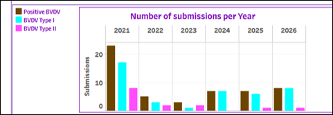

The Interactive Animal Pathogen Dashboards (IAPD) provides trends in key cattle pathogens using Animal Health Laboratory (AHL) submission data. Dashboards report total submissions, positive results, and percent positive by week, month, quarter, and year.
Available bovine dashboards include: Bovine abortion, Bovine viral diarrhea virus (BVDV), Influenza A virus, Mycoplasma bovis, Salmonella Dublin (Bulk), Salmonella Dublin (Individual), and Salmonella in cattle.
Veterinary clinics can request a login by contacting Tanya Rossi (rossit@uoguelph.ca).
HPAI in Dairy Cattle in the U.S.
In 2026, 89 cases of HPAI in dairy cattle have been reported in the United States, as of September 24, 2026. This is substantially fewer than in previous years, with 171 cases reported in 2025 and 917 detections in 2024. Following a relatively quiet period during mid-summer, late-August updates confirmed a small number of new cases, including four affected herds in Texas and two in Idaho. Earlier in June, clusters of cases were identified in Idaho and Utah. It is important to note that previously affected dairies that subsequently become re-infected are not counted a second time in USDA reporting. As a result, the total number of herds affected may be underrepresented in USDA summary statistics.
Theileria orientalis Ikeda Distribution in the U.S.
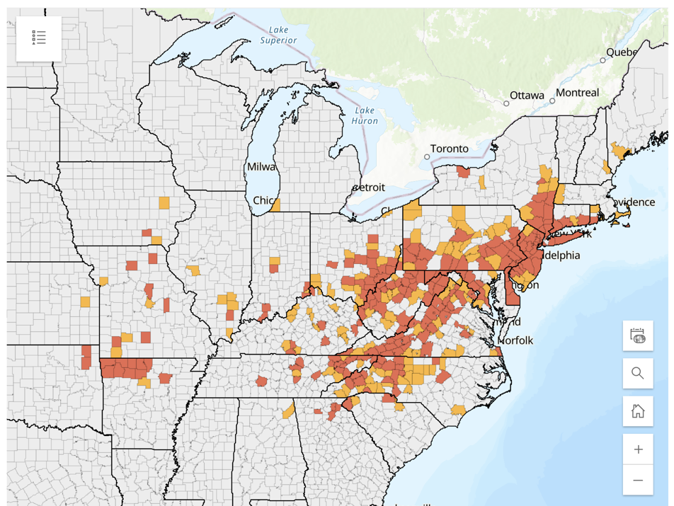
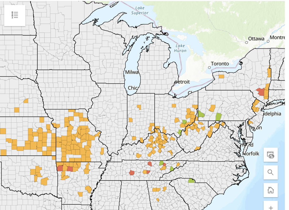
The University of Tennessee released updated maps on the distribution of Asian longhorned tick (Haemaphysalis longicornis) and Theileria orientalis Ikeda as of August 2026. Theileria orientalis Ikeda infection can cause anemia, lethargy, reduced milk production, weight loss, abortion, and, in severe cases, death.
Given the potential for introduction of tick-borne pathogens, veterinarians should consider recommending quarantine, careful inspection for ticks, and PCR blood testing of newly acquired animals originating from the U.S.
Other Resources:
OAHN Factsheets: Factsheet: Bovine Theileriosis and Factsheet: Asian Longhorned Tick
Interesting article from Bovine Veterinarian: From Dead Heifers to a Herd-Wide Discovery
This investigation highlights the importance of considering Theileria orientalis Ikeda in cattle with
unexplained anemia or mortality, and the need for continued tick surveillance and control.
Case Report: E. coli septicemia in group of Holstein-Angus cross calves
Contributed by OAHN member Dr. Daniel Pecoskie
A producer in southwestern Ontario was experiencing an increase in mortality in 450-600lb Holstein-Angus cross stocker steers and requested a veterinarian perform postmortem examination of three recently deceased calves. The calves were described as having developed subtle lameness and general stiff, shortened gait on all four limbs. The stiffness progressed over 3-4 days despite treatment with systemic antibiotics, and steers went down in lateral recumbency, with paddling of limbs and opisthotonus before being humanely euthanized or found acutely dead in pen. The symptoms coincided with recent changes to the vaccine protocols, as well as increased stocking density follow transport between barns.
The calves are sourced in groups of 75-head at 2- weeks of age from Quebec and transported to southwestern Ontario where they are raised in a continuous flow system. Calves start in hutches at a farm across the road. They are vaccinated on arrival with a three-way modified live viral intranasal vaccine (which is boostered at 3-weeks) and fed milk and calf starter for 7-weeks. From there, they are weaned and moved to a coverall barn next to the hutches, where they are backgrounded for another 7-weeks. Calves are vaccinated with a subcutaneous 5-way MLV + 2-way bacterin vaccine at 8-weeks and 28-weeks post arrival. Subsequently, calves are moved to the affected operation where they are processed on arrival with tulathromycin, at around 450lb body weight. Recently, the vaccine protocol has changed to include a subcutaneous mycoplasma vaccine at 2-weeks and 6-weeks post-arrival. In the last two groups, totalling 150 calves, seven animals died at the affected barn after developing a stiff gait that progressed to lateral recumbency.
Postmortem findings found petechial hemorrhages over the surface of filter organs, and one calf showed signs of acute bronchointerstitial pneumonia affecting 30% of cranioventral lung lobes on the left and right side. A second calf showed fibrin covering the pericardial sac, mild pleural effusion, with fibrin also adhered to 10-inch section of serosal surface of proximal jejunum. The mesenteric lymph nodes in this region were enlarged, and the vessels of the mesentery were shocked and prominent. Bacterial septicemia leading to signs of arthritis and neurologic symptoms was suspected. Fresh and fixed tissues, and joint fluid were submitted to AHL Guelph for histopathology, and bacterial culture and sensitivity.
Histopathology of assorted tissues found a range of findings including multi-organ congestion, acute pulmonary edema with intralesional bacteria, mild pleuritis and serositis, fibrinosuppurative bronchointerstitial pneumonia and mild acute embolic hepatitis, which supported the working diagnosis of bacterial septicemia.
Bacterial culture and sensitivity of two submitted joint fluid samples collected aseptically from the stifle joints cultured 1+ and 4+ E.coli respectively, and the fresh lung sample submitted cultured 1+ E.coli. Salmonella Dublin PCR of the fresh lung tissue tested negative.