
Ontario Animal Health Network (OAHN) Poultry Expert Network Quarterly Veterinarian Report
Enterococcus cecorum in broiler chickens: An ongoing concern
Emily Martin, Animal Health Laboratory, University of Guelph, Guelph, ON.
Prior to 2002, E. cecorum was considered part of the normal intestinal flora in avian species (e.g. turkeys, Muscovy ducks, pigeons, canaries) and mammals (e.g. horses, cattle, pigs, dog, cat) but identification of pathogenic strains has had a significant impact on broiler health and production. Pathogenic strains of E. cecorum were identified in chickens starting with outbreaks in Scotland and the Netherlands (2002) then progressing to other countries (e.g. Canada, USA, Iran, South Africa). Broiler production is impacted by flocks experiencing increased morbidity (up to 35%), retarded growth, increased mortality (up to 15%), and increased condemnation rates (up to 10.6%).
Clinical presentation:
E.cecorum infection can be described as two stages of disease. Initially, there may be no observable clinical signs (asymptomatic or subclinical) during the initial infection in 2-3 week old broilers or there may be clinical signs of depression (i.e. ruffled feathers, closed eyes). As infection progresses, bacterial sepsis can cause lesions of hepatitis, pericarditis and splenomegaly as well as bacteria localizing in bones or joints resulting in increased mortality, decreased mobility and lameness. When bacteria localize in the free thoracic vertebrae (FTV), older broilers (over 3 weeks) can develop bilateral paralysis where birds can be found hock sitting with their legs extended forward. Mortality is usually higher at this stage due to culling, dehydration and starvation.
Postmortem lesions:
On postmortem examination, early infection can be identified through lesions of pericarditis, hepatitis and splenomegaly. These lesions are similar to other bacterial infections; therefore, bacterial culture is recommended. In later infections, joint lesions (i.e. synovitis and arthritis of the coxofemoral, stifle, and hock joints) have accumulation of creamy yellow fluid and sometimes yellow caseous material resulting in joint swelling. Bone lesions (i.e. spondylitis, osteomyelitis) can be identified in the FTV, proximal femur, or proximal tibia. Bacteria are thought to locate in these lesions due to anatomy and small microfractures created due to shearing forces of movement. Lesions in the free thoracic vertebra and adjacent vertebral bodies cause necrosis and granuloma (or abscess) formation that expands dorsally to cause spinal cord compression resulting in bilateral paralysis. In severe cases, this vertebral mass expands ventrally into the celomic cavity and creates a localized enlargement of the spinal column that can be seen around the caudal lungs/cranial kidneys.
Diagnosis:
As lesions of E. cecorum septicemia are similar to other infectious bacteria, bacterial culture is required for confirmation. It is possible to culture E. cecorum from the FTV and femoral heads without gross lesions. Virulence among pathogenic strains varies significantly. In multiple studies it was found that commensal (E. cecorum from normal flora) isolates have higher diversity whereas pathogenic isolates originating from different countries group phylogenetically, primarily belong to one clade, and are distinct from the commensal poultry isolates. There are few virulence factors identified in both the commensal and pathogenic strains of E. cecorum.
Pathogenesis and transmission:
The pathogenesis and transmission of E. cecorum is still not well understood. It is thought that pathogenic strains colonize the intestines, allowing rapid spread throughout a flock (primarily fecal-oral transmission). However, due to increased intestinal permeability, potentially related to intestinal damage, the bacteria move out of the intestines to other organs including the heart, spleen, bones, and cartilage to cause systemic infections. Some of these sites (i.e. heart, cartilage) make it difficult for antibiotics to reach the required drug concentration, potentially contributing to persistent infections. Pathogenic strains can also be multi-drug resistant, but resistance patterns do vary between isolates making antimicrobial sensitivity testing important. Other potential routes of infection include through the skin or through air sacs that extend into pneumatic bone.
Survivability:
It is also known that Enterococcus spp. (including E. cecorum) survives well in the environment, potentially being a source of repeated infections within a barn. Research has shown that E. cecorum survives longer in litter than in dust; there is longer survival at lower temperatures, and there is longer survival at lower relative humidity. Pathogenic strains showed a longer survival time than commensals under similar conditions.
It is known that E. cecorum infections can be repeated as seen in subsequent production cycles in the same barn. Phylogenetic comparison of isolates of E. cecorum from the same farm over multiple production cycles revealed either long-term persistence or repeated introductions. Pathogenic strains are more persistent in the environment and can survive cleaning and disinfection.
Detection of E. cecorum in the environment is difficult, and research investigations have used PCR rather than culture methods. While PCR can detect both viable and non-viable organisms, it is still notable that when the barn environment was sampled, it was found that there were increased positive samples toward the end of the production cycle. Increased positive sampling locations include air exhausts, boot covers, carcass buckets, drinking nipples, drain holes, floor cracks and feces. After cleaning and disinfection, there were fewer environmental samples that tested positive, however, since some environmental samples still tested positive for E. cecorum after cleaning and disinfection points to the environment as a potential source of infection between flocks.
Conclusion:
Currently, our understanding of E. cecorum is still evolving in avian species. Further research into transmission and sources of infection will aid in developing prevention and management strategies that currently rely on strict biosecurity, thorough cleaning and disinfection as well as feed and water quality management.
References:
- 2023 ACPV Workshop. Enterococcus: An Old Dog with New Tricks. March 12, 2023 Sacramento, CA. https://aaap.memberclicks.net/assets/ACPV_Workshops/2023_Workshop/2023_ACPV%20Workshop%20Program.pdf
- Cordoni, G., Genomic characterisation of Enterococcus cecorum isolated from broiler chickens in the United Kingdom, Poultry science, 2026 https://doi.org/10.1016/j.psj.2026.107243.
- Grund A., Rautenschlein S. and Jung A. Tenacity of Enterococcus cecorum at different environmental conditions. J Appl Microbiol. 2021;130: 1494-1507. https://doi.org/10.1111/jam.14899
- Jung, A., et al. A Review of Enterococcus cecorum Infection in Poultry. Avian Diseases. 2018;62(3), 261–271. https://www.jstor.org/stable/26537546
- Laurentie, J., et al. Comparative Genome Analysis of Enterococcus cecorum Reveals Intercontinental Spread of a Lineage of Clinical Poultry Isolates. 2023;8(2):1-21. https://doi.org/10.1128/msphere.00495-22
- Schreier J, Rautenschlein S, Jung A. Different virulence levels of Enterococcus cecorum strains in experimentally infected Meat type chickens. PLoS ONE. 2021;16(11): e0259904. https://doi.org/10.1371/journal.pone.0259904
- Tessin, J., et al. (2024). Detection of Enterococcus cecorum to identify persistently contaminated locations using faecal and environmental samples in broiler houses of clinically healthy flocks. Avian Pathology, 2024;53(4): 312–320. https://doi.org/10.1080/03079457.2024.2334682
- Wijetunge DS, et al. Fingerprinting of poultry isolates of Enterococcus cecorum using three molecular typing methods. JVDI. 2012;24(6):1166-1171. https://doi.org/10.1177/1040638712463563
Poultry Veterinarian Survey Highlights – Q2 2026 (Mar-May 2026)
Broilers
Practitioners
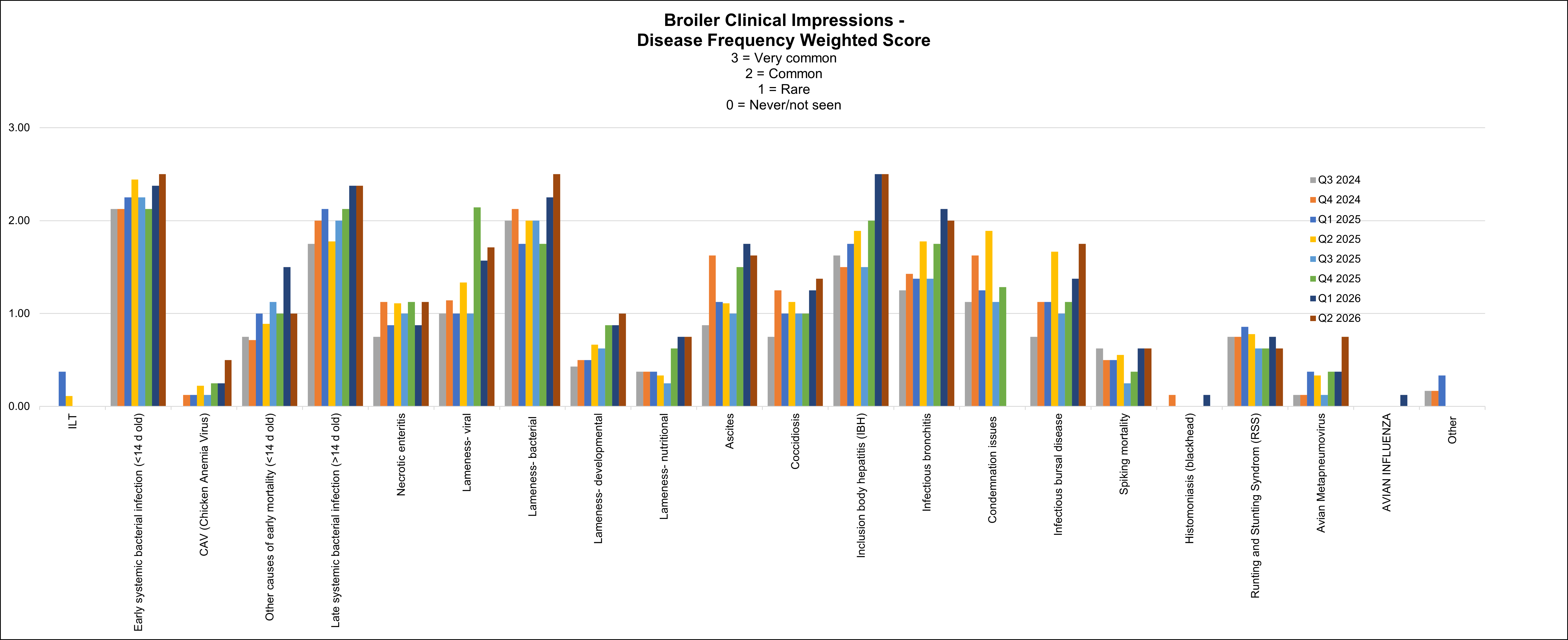
Stable to increased conditions: early and late systemic bacterial infection due to E. coli and E. cecorum infections, lameness bacterial origin, lameness of viral origin (reovirus related), inclusion body hepatitis (IBH) and avian metapneumovirus (AMPV).
Stable to equally slightly increased or decreased conditions: coccidiosis, other causes of early mortality (dehydration, IBH and necrotic enteritis).
Stable conditions: Infectious laryngotracheitis (ILT), chicken anemia virus (CAV), ascites, infectious bursal disease (IBVD), histomoniasis, spiking mortality and avian influenza.
Stable to decreased conditions: runting stunting syndrome (RSS), infectious bronchitis (IBV), necrotic enteritis, lameness developmental and nutritional causes.
Comorbidities described with AMPV infections were bacterial infections (E. coli most common) with late break of infection, IBV, increased DOA and excessive condemnation rate on broilers presented to slaughter due to respiratory conditions.
A range between 10-45 % of E. coli isolates were described by practitioners to be resistant to trimethoprim sulpha (TMS) in broilers.
Below graph depicts the frequency of the conditions seen in the flocks. The most frequent 3 conditions seen this quarter were: early systemic bacterial infection (<14 d old), lameness (bacterial) and Inclusion body hepatitis (IBH).
AHL
Similar number of cases (from previous quarter):
– Early systemic bacterial infection (< 14 days old) – All cases of septicemia had E. coli isolated in pure culture or with E. cecorum.
– Other causes of early mortality (< 14 days old) – Urate nephrosis and mycotic pneumonia / airsacculitis / meningoencephalitis.
– Lameness developmental (tibial dyschondroplasia).
-Necrotic enteritis.
Increased number of cases (from previous quarter):
– Late systemic bacterial infection (> 14 days old) – E. coli and E. cecorum isolated in either in pure culture or in combination.
– Pododermatitis.
– Lameness bacterial (Primarily diagnosed on histology) – E. coli isolated in pure culture or in combination with E. cecorum or P. aeruginosa. Eleven cases of osteomyelitis had E. coli isolated in pure culture or with E. cecorum.
– Lameness viral (Reovirus suspicious, primarily diagnosed on histology).
– Inclusion body hepatitis (FAdVE/FAdVAC, FAdVD/FAdVAC, FAdVE/FAdVD/FAdVAC, FAdV08b_AHL_16-049095_ON and FAdV08b_AHL_18-057921_ON and FAdV11_USA_1047).
– Infectious bursal disease (PCR positive field strains: USA PA105-2014, British Columbia 15-062782, USA Del-E and USA Del-A-66-Indiana-2014. Vaccine strains: UK Faragher52-70).
– Wooden Breast (Processing samples).
Decreased number of cases (from previous quarter):
– Lameness nutritional (rickets).
– Lameness viral (Reovirus confirmed: ON_variant_H_18-008168, ON_variant_A_12-073195, ON_variant_G_16-102019, SK-R10, Alberta 17-0025 and Pennsylvania 9552).
– Coccidiosis (small intestine or ceca).
– Infectious bronchitis (IBV – pathology cases and PCR positive) (Field strains: IBV_DMV_ON_21-017385. Vaccine strains: IBV_Conn, IBV_Mass-MA5, USA Mass-AHL 21-008165 and USA GA08-IBron).
– Avian metapneumovirus (Type B, Turkey ONT 24-093127-0001).
– RSS – Confirmed.
– RSS – Suspect.
No cases diagnosed this quarter:
– Spiking mortality.
– Botulism.
– ILT.
– Dead on arrival (DOA).
Other diagnostic findings:
Sodium toxicosis, bacterial pneumonia (S. Typhimurium / E. coli / E. cecorum, E. coli / E. cecorum), uveitis, myopathy (E. coli / E. cecorum), necrotizing enteritis, typhlitis (heterophilic), cellulitis and M. synoviae.
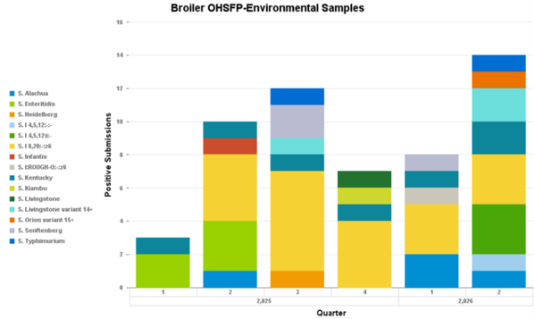
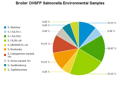
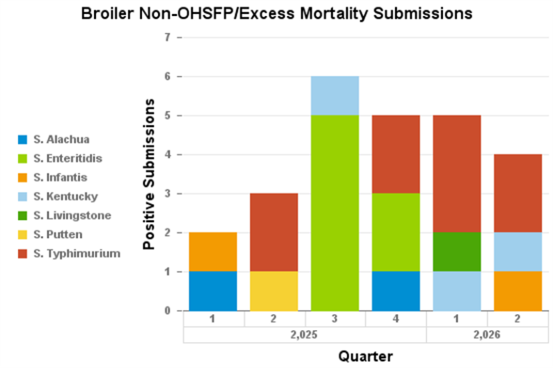
Salmonellosis (Salmonella isolation)
Slaughter data
Chickens slaughtered and carcass condemnation (December 2025- February 2026)
Condemnation conditions
Provincial slaughter: top 3 conditions of condemnation- ascites (0.57%), peritonitis (0.46%) and air sacculitis/CRD (0.32%).
Federal slaughter (CAHSS data): top 3 conditions- subcutaneous, liver and abdominal oedema
Broiler-Breeders
Most of the diseases were stable for this quarter with an increase in AMPV cases and decrease in IBV detections compared with last quarter.
The critical issue the producers are dealing with this summer is heat stress related morbidity and mortality, especially in flocks located in barns not equipped with tunnel ventilation systems. In response, producers are using some interventions that include adding electrolytes with the drinking water, feed reduction (this measure negatively impacts production), running cooling down ventilation during night time and in extreme cases spraying water on the roof of the barn.
In-lay bacterial septicemia and bacterial lameness were attributed to E. coli, E. cecorum and S. aureus.
Stable conditions: fowl cholera, CAV, lameness developmental/nutritional/ viral, necrotic enteritis, fowl pox, ILT and avian influenza.
Stable to slightly increased conditions: early bacterial infections, other causes of early mortality (<14 days old), lameness bacterial, coccidiosis, aggression and cannibalism (feather licking), multi-drug-resistant E. coli (> 3 drugs) and pre-lay (<20 weeks) morbidity/mortality.
Stable to slightly increased conditions: early bacterial infections, other causes of early mortality (<14 days old), lameness bacterial, coccidiosis, aggression and cannibalism (feather licking), multi-drug-resistant E. coli (> 3 drugs) and pre-lay (<20 weeks) morbidity/mortality.
Stable to increased conditions: AMPV with clinical signs in affected flocks related to IBV and E. coli infections, drop in egg production, in-lay septicemia, and egg peritonitis.
It was also noted that in vaccinated flocks egg production drop was not observed and mortality seems to be reduced compared to non-vaccinated flocks.
Stable with equally increased or decreased conditions: IBV – decreased production/abnormal eggs, mycoplasmosis, histomoniasis and in-lay bacterial septicemia mostly seen after the peak.
Stable to slightly decreased conditions: disease related hatchability issues attributed to AMPV infections, IBV – sudden spike in mortality and mycoplasmosis (M. synoviae).
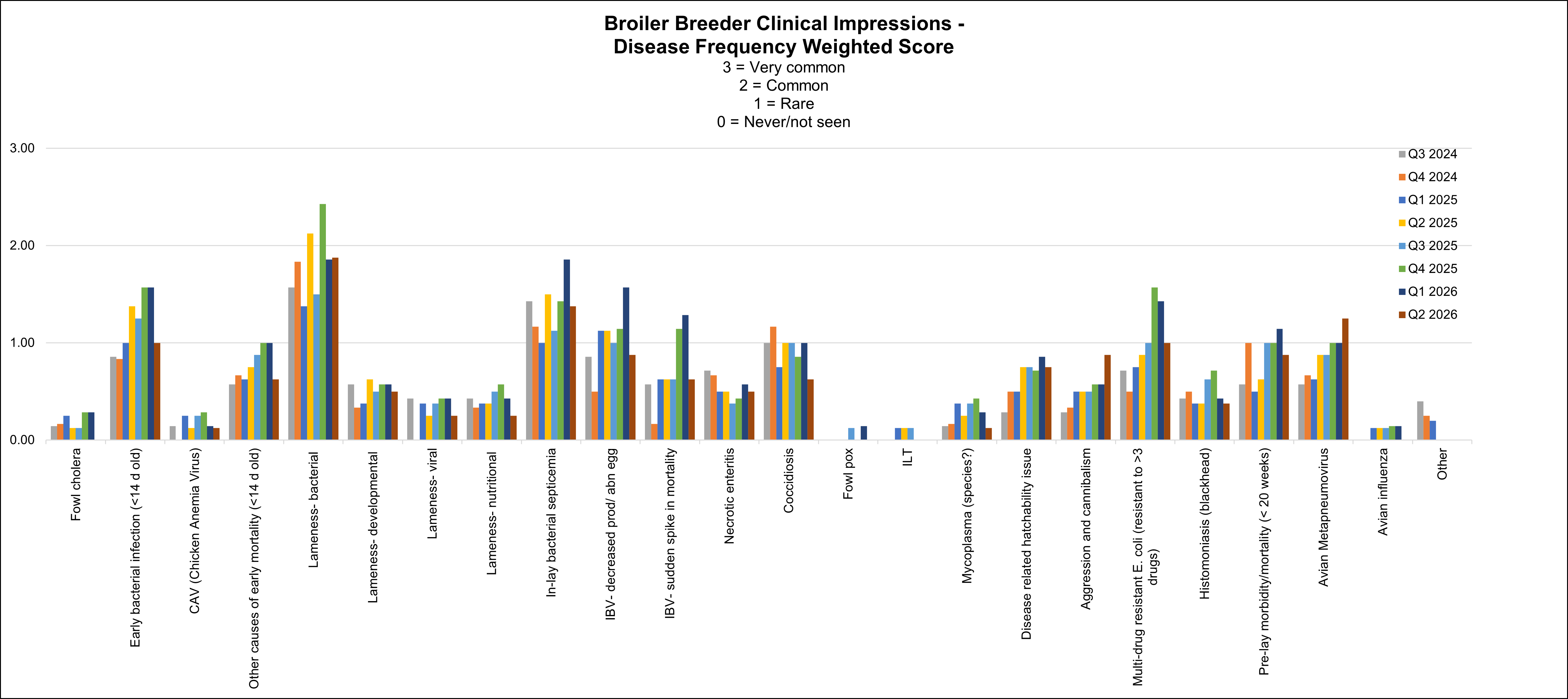
Below graph depicts the frequency of the conditions seen in the flocks. The most frequent conditions seen this quarter were: lameness (bacterial), in-lay bacterial septicemia and AMPV.
AHL
Fowl cholera (Pasteurella multocida) – Occasionally isolated from lameness cases from vaccinated flocks.
Similar number of cases (from previous quarter):
– Prelay morbidity/mortality (<20 weeks) – S. aureus was isolated in pure culture or with E. coli and E. cecorum. E. coli and E. cecorum were isolated together. E. cecorum was also isolated with P. multocida. One other diagnosis was CAV_On_AHL13-012821-0006.
– Other causes of mortality (> 14 days old) – Urate nephrosis and tongue / beak necrosis.
– Bacterial pneumonia – E. coli and E. cecorum were isolated together as well as with G. anatis.
– Avian metapneumovirus (Type B).
– IBV (decreased productivity/abnormal eggs/sudden spike in mortality) – (Field strain: None. Vaccines: IBV_Conn, IBV_Mass-MA5 and USA GA08-IBron).
– White Chick Syndrome.
Increased number of cases (from previous quarter):
– Inlay bacterial septicemia – E. coli and S. aureus were each isolated in pure culture. E. coli was also isolated with either S. aureus or E. cecorum. E. coli and E. cecorum were isolated with S. aureus, G. anatis, ORT, S. aureus and P. aeruginosa, or C. septicum and C. perfringens.
-Cellulitis/Dermatitis.
-Lameness trauma – Tendon rupture.
-Pododermatitis – E. coli, E. cecorum and S. aureus isolated together. Avibacterium spp. Isolated in pure culture.
– Lameness bacterial – S. aureus and E. coli were each isolated in pure culture as well as together. S. aureus was also isolated with E. cecorum or both E. coli and E. cecorum. P. multocida was isolated with either E. coli, S. aureus and E. coli, or S. aureus and E. cecorum. One case of osteomyelitis had E. coli, E. cecorum and S. aureus isolated together.
– Lameness viral – Reovirus (Identified on PCR).
– Necrotic enteritis (C. perfringens isolated).
– Intestinal parasitism – Histomonas and nematodiasis (Ascarids, Heterakis).
Decreased number of cases (from previous quarter):
– Early bacterial infection (<14 days old) – Cases of septicemia had E. coli isolated in pure culture.
– Coccidiosis – Identified in the small intestine (E. necatrix) or ceca (E. tenella).
– Amyloidosis.
No cases diagnosed this quarter:
– Lameness nutritional (rickets).
– Lameness developmental.
– Fowl pox.
– Mycoplasmosis.
– Other causes of inlay mortality.
– Urate nephrosis/visceral urates/articular urates.
Other diagnostic findings:
Gangrenous dermatitis (E. coli, S. aureus / E. coli, S. aureus / E. coli / C. perfringens, S. aureus, / E. coli / E. cecorum, S. aureus / E. coli / E. cecorum / C. perfringens), lymphoid neoplasia, corneal ulceration / conjunctivitis / uveitis, sinusitis (E. coli / E. cecorum), necrotizing stomatitis, liver hemorrhage, yolk peritonitis, cystic oviduct, salpingitis (E. coli, E. coli / E. cecorum, E. coli / E. cecorum / S. aureus) and IBH (FAdV08b_AHL_18-057921_ON and FAdV11_USA_1047).
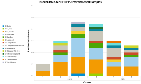
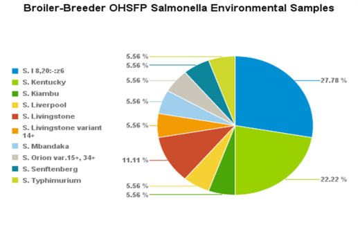
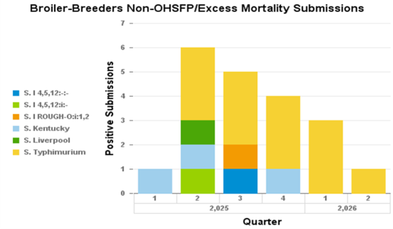
Salmonellosis (Salmonella isolation):
Layers
Veterinarians continue to report challenges of the health status in the flocks since September 2025, with increasing cases of peritonitis/salpingitis (0.5-1% of the birds in the flock). AMPV ELISA positive testing on the submitted samples increased from 70 % during last quarter to 80% this quarter. There was also one detection of IBV GA08 variant strain in a layer flock that never received a specific GA08 vaccine.
Practitioners
Stable to increased conditions: bacterial peritonitis/salpingitis early in the lay period (E. coli), and AMPV. Clinical signs seen with AMPV cases: E coli septicemia, salpingitis, suspecting mycoplasmosis but have not been tested; increased mortality just before or after coming into lay; peritonitis; salpingitis; not coming into or drop in production with no egg abnormalities noted; reduced feed intake; IBV and AMPV seem linked.
Stable to slightly decrease in S. enteritidis detection.
Stable conditions: osteoporosis, other causes of mortality, early systemic bacterial infection, focal duodenal necrosis, IBV respiratory issues, ILT, mycoplasmosis, coccidiosis, hysteria, aggression, and cannibalism, multi drug resistant E. coli (resistant to > 3 drugs but widely susceptible to tetracycline), histomoniasis and avian influenza.
Stable with equally increased or decreased conditions: necrotic enteritis (not NE necessarily, but Clostridium enteritis).
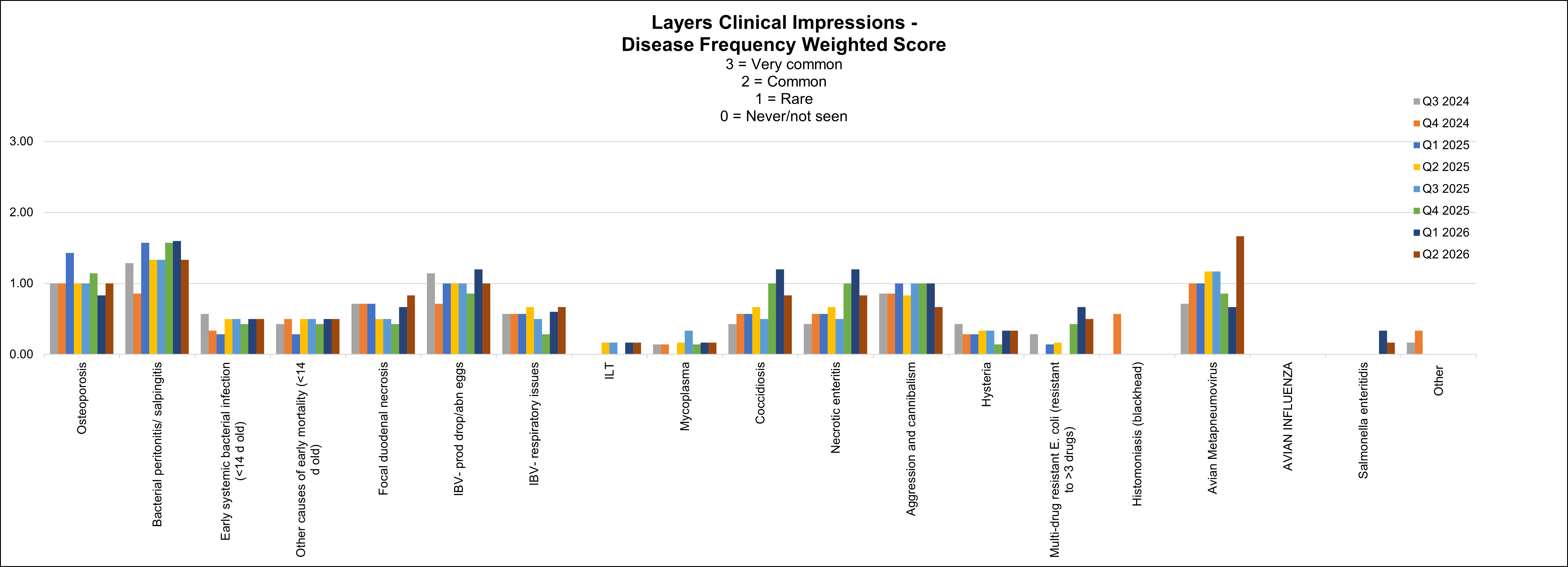
Below graph depicts the frequency of the conditions seen in the flocks. The most frequent conditions seen this quarter were: AMPV, bacterial peritonitis/ salpingitis and IBV- prod drop/abnormal eggs.
AHL
Similar number of cases (from previous quarter):
– Coccidiosis (Small intestine).
Increased number of cases (from previous quarter):
– Early mortality (<14 days old, starve outs).
– Osteoporosis.
– Necrotic enteritis (C. perfringens).
– Avian metapneumovirus (Type B).
Decreased number of cases (from previous quarter):
– Prelay mortality (<20 weeks old) – Disseminated fungal granulomas.
– In-lay mortality (>20 weeks old) – Septicemia (E. coli).
– Leg issues – Bacterial (tenosynovitis/arthritis).
– Infectious bronchitis (IBV – production drop/egg abnormalities/ respiratory) – Field strains: None. Vaccine strains: IBV_Conn, IBV_Mass-MA5, USA GA08-IBron and USA DE-072.
No cases diagnosed this quarter:
– Early mortality (<14 days old, omphalitis).
– Early mortality (other).
– Leg issues – Trauma, bone fracture.
– Leg issues (other).
– Focal duodenal necrosis (FDN).
– Intestinal parasitism (other).
– Aggression/Hysteria.
– ILT.
– Mycoplasma.
– Elevated DOA.
Other diagnostic findings:
Salpingitis (E. coli, E. coli / E. cecorum / S. suis), corneal erosion / conjunctivitis / keratitis / iridocyclitis / uveitis, peritonitis / oophoritis, amyloidosis, vaccine reaction (systemic), pneumonia, conjunctivitis / tracheitis, vaginal prolapse, vent pecking / cannibalism, Marek’s disease (eyes) and Fowl Adenovirus (FAdVAC/FAdVE).
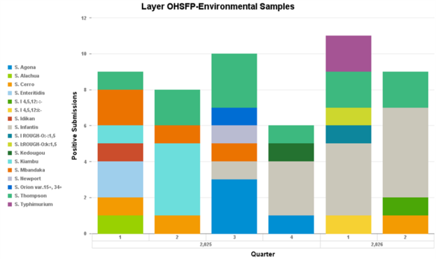
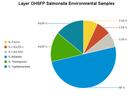
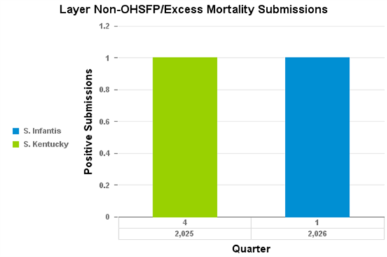
Salmonellosis (Salmonella isolation)
Turkeys
Practitioners
Turkey flock health conditions were mostly stable this quarter. AMPV infections continue to be detected in the flocks and are linked with late bacterial infection resulting in pneumonia and specific E.coli fibrino-purulent lesions affecting air-sacs, liver, pericardium and peritoneum.
Stable conditions: fowl cholera, erysipelosis , other respiratory diseases (bacterial pneumonia and AMPV), necrotic enteritis, coccidiosis, round heart, mycoplasmosis, aggression, and cannibalism.
Stable to increased conditions: late systemic bacterial infection (E. coli) and AMPV.
Clinical signs and pathology seen in flocks affected by AMPV infection:
- severe systemic bacterial infections mostly attributed to E. coli infection;
- snicking/depression in birds with upper respiratory clinical signs;
- cannibalism and head pecking;
- acute death and increased mortality due to respiratory signs;
- peritonitis, pericarditis and pneumonia.
Stable to slightly increased conditions: ORT, enteritis, reovirus tenosynovitis, multi drug resistant E. coli (resistant to > 3 drugs), early systemic bacterial infection (E. coli).
Stable to slightly decreased conditions: other causes of mortality, mycotic respiratory disease, histomoniasis and avian influenza.
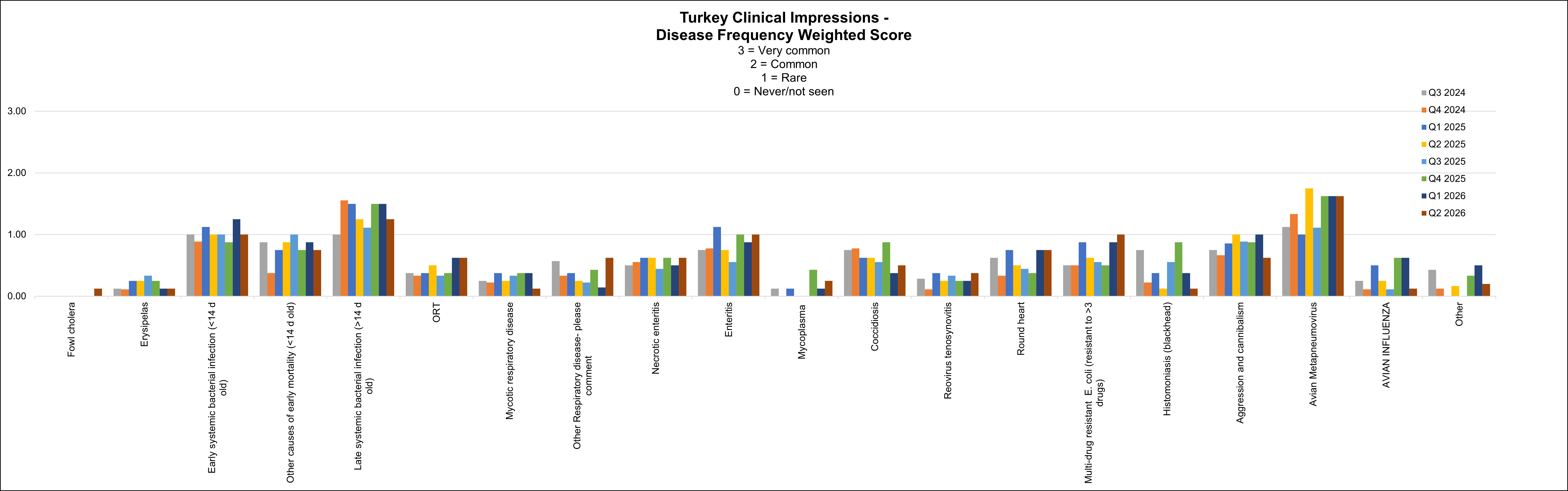
Below graph depicts the frequency of the conditions seen in the flocks. The most frequent 3 conditions seen this quarter were: AMPV, late systemic bacterial infection (>14 d old) and enteritis.
AHL
Similar number of cases (from previous quarter):
None.
Increased number of cases (from previous quarter):
-Avian metapneumovirus – Type B (Turkey ONT 24-093127-0001) and Type A (vaccine?).
– Rickets.
Decreased number of cases (from previous quarter):
– Late systemic bacterial infection (>14 d old) – E. coli and P. aeruginosa isolated together.
– Tibial dyschondroplasia.
– Reovirus – Histology.
No cases diagnosed this quarter:
– Fowl cholera (Pasteurella multocida).
– Erysipelas.
– Other causes of early mortality (<14 days old, starve out / dehydration).
– Early systemic bacterial infection (<14 days old)
– ORT (Ornithobacterium rhinotracheale).
– Bacterial pneumonia.
– Mycotic respiratory disease.
– Enteric disease.
– Coccidiosis/ Parasitism.
– Mycoplasma.
– Necrotic enteritis.
– Histomoniasis.
– Salmonellosis.
– Condemnations.
– DOA.
Other diagnostic findings:
Blepharitis / keratoconjunctivitis / iridocyclitis (E. coli, S. aureus), chondrodystrophy, bacterial osteomyelitis (E. coli, P. aeruginosa), tenosynovitis, aorta (cartilaginous metaplasia) and myodegeneration.
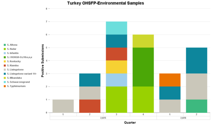
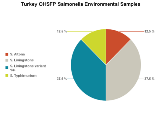
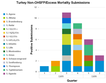
Salmonellosis (Salmonella isolation)
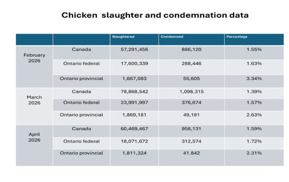
Slaughter data (February-April 2026)
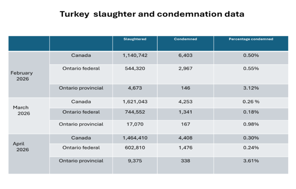 Condemnation conditions:
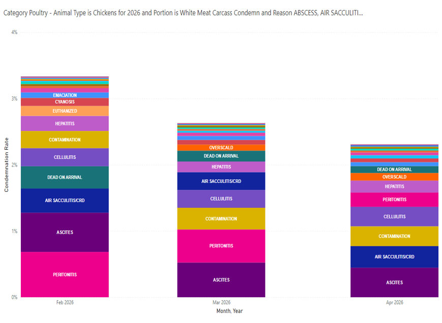
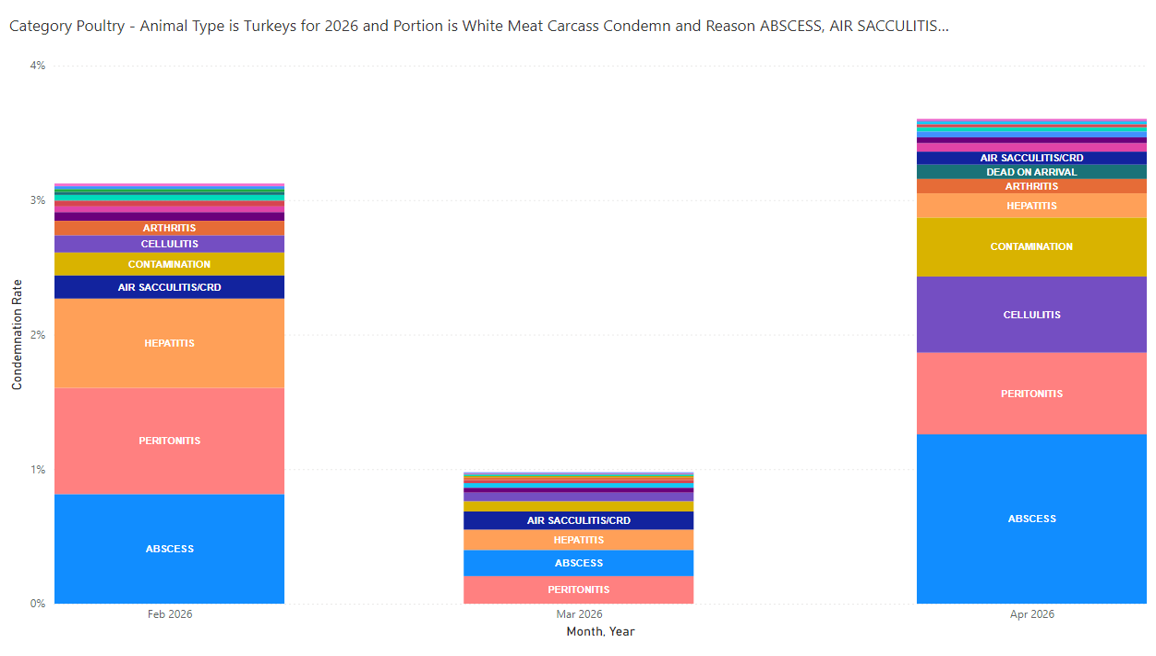
Provincial slaughter– top 3 main conditions listed for this quarter were: abscesses (0.61%), peritonitis (0.41%) and hepatitis (0.24%).
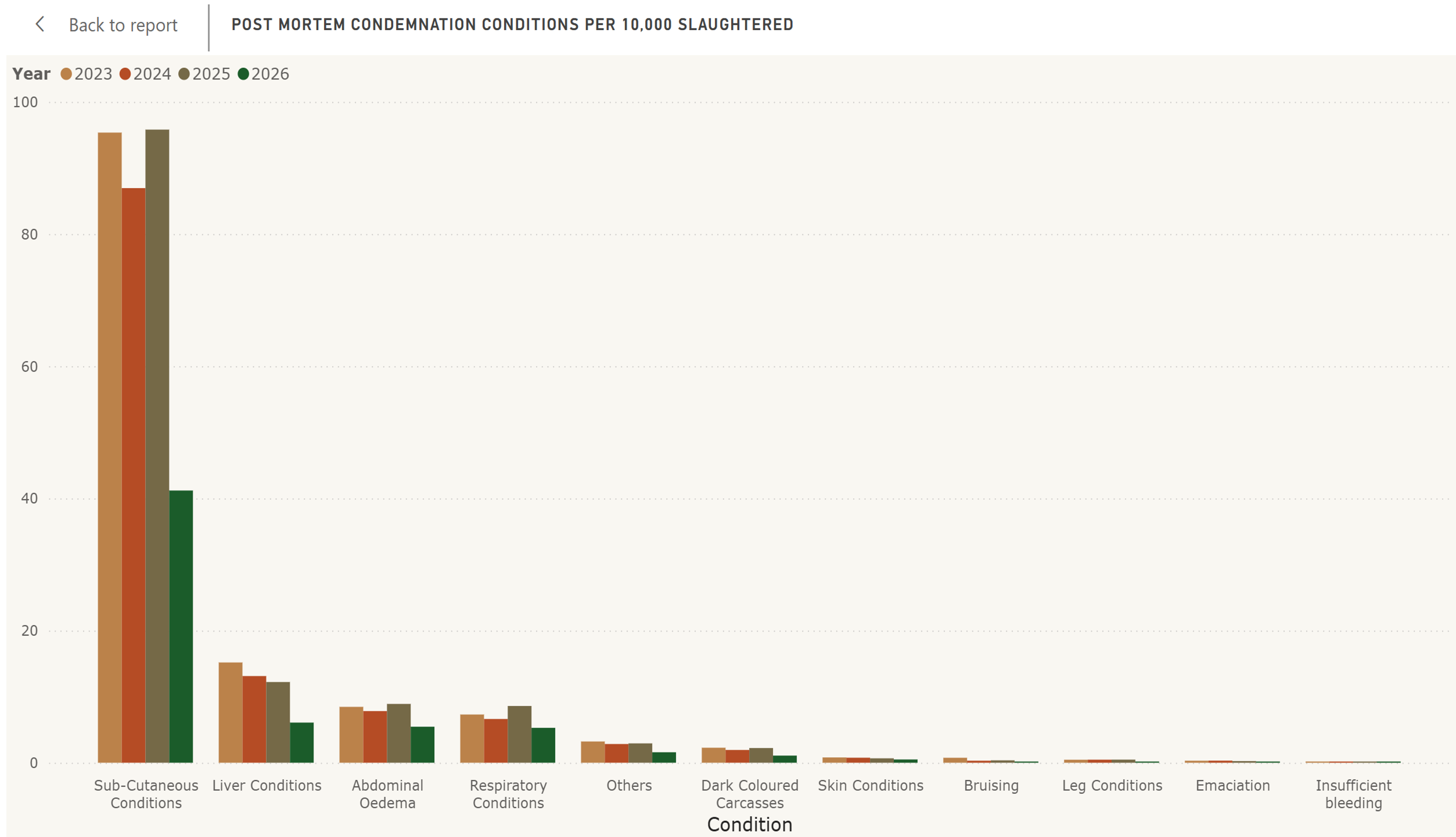
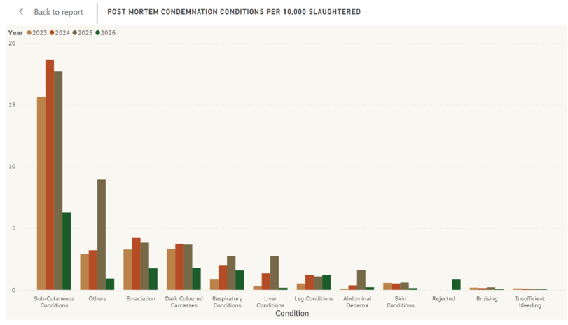
Federal slaughter (CAHSS dashboard)
Top 3 conditions listed for this quarter were: subcutaneous, dark coloured carcasses and emaciation.
Small Flock
Chickens:
AHL: Diagnoses at AHL included intestinal parasitism (coccidiosis), Marek’s disease, neoplasia (lymphoid neoplasia, adencarcinoma), peritonitis/salpingitis/celomitis, bacterial septicemia, bronchopneumonia (suspect fungi), FLHS, acites, necrotizing stomatitis and esophagitis, IBV (suspicious histology), diarrhea and mucosal hyperplasia/ballooning degeneration.
Gamebird/Ratites:
AHL: One Golden pheasant submitted the quarter.
1) Ligament arthropathy, tendinopathy – hock.
Waterfowl:
AHL: Four ducks submitted this quarter.
1) Duck – Testicular neoplasia with metastases.
2) Runner Duck – Adenocarcinoma, salpingitis.
3) Peking Duck – Septicemia (suspect).
4) Ducks (at processing) – Lymphoid neoplasia.
Turkey:
AHL: No cases submitted this quarter.
Pigeon:
AHL: One case submitted this quarter (2 weeks old).
1) Crop mycosis, S. Typhimurium septicemia.
Practitioners: Information provided with the clinical impression in chickens:
Frequency of conditions seen in practice:
Very Common : Ectoparasitism.
Common : Intestinal parasitism, Marek’s disease, other neoplasia, GI impaction, trauma, pododermatitis, peritonitis/salpingitis, ascites, Mycoplasma, other (feather-pecking).
Rare : Vent trauma, bacterial septicemia, pneumonia/airsacculitis, FLHS, urate nephrosis/ visceral urates/articular urate deposits, ILT, toxin.
Not seen : Histomoniasis, erysipelas, and AMPV.
Changes since last quarter: stable conditions: ectoparasitism, other neoplasia, vent trauma, peritonitis/salpingitis, bacterial septicemia, pneumonia/airsacculitis, FLHS, urate nephrosis/ visceral urates/articular urate deposits, ILT; increased cases of pododermatitis, other (feather-pecking), decreased cases of intestinal parasitism, Marek’s disease, GI impaction, trauma, ascites, toxin, Mycoplasma.
Poultry research from Ontario and beyond
- Transmission dynamics of highly pathogenic avian influenza virus at the wildlife-poultry-environmental interface: A case study – PubMed
- One Health, Many Realities: Navigating Collaboration for Avian Influenza Surveillance and Response | One Health Cases
- Differentiation of Pathogenic from Nonpathogenic Enterococcus cecorum Field Isolates Based on the Detection of the cpsO Gene and Its Association with Macroscopic Lesions in: Avian Diseases Volume 70: Issue 2 | Avian Diseases
- Comprehensive Pathologic Evaluation of Histomonas meleagridis Field Isolates in Target (Cecum and Liver) and Primary Lymphoid (Bursa and Thymus) Organs in Turkeys in: Avian Diseases Volume 70: Issue 2 | Avian Diseases
Thank You! We thank the following poultry veterinarians who completed the veterinary survey:
Dr. Tim Abolarin, Dr. Elizabeth Black, Dr. Daniella Di Pirro, Dr. Fernando Salgado-Bierman, Dr. Shahbaz Haq, Dr. Elana Huong, Dr. Anastasia Novy, Dr. Mike Petrik, Dr. Joanne Rafuse, Dr. Nahal Ramezani, Dr. Ben Schlegel, Dr. Hillary Schramm, Dr. Chanelle Taylor, Dr. Brenna Tuer, Dr. Alex Weisz, and Dr. Jessalyn Walkey. Small Flock: Dr. Amari Patel.