
Ontario Animal Health Network (OAHN)
Small Ruminant Expert Network
Report
Animal Health Laboratory Summary Oct 2025-March 2026
Ovine: 40 postmortem cases and 46 histopathology cases from both laboratory and on-farm postmortems/biopsy were submitted to the AHL and OVC-PBI from October 1, 2025, to March 31, 2026. A total of 28 of these cases had both postmortem and histology performed; comprising 70% of total postmortems submitted.
Caprine: 38 postmortem and 36 histopathology cases from both laboratory and on-farm postmortems/biopsy were submitted to the AHL and OVC-PBI from October 1, 2025, to March 31, 2026. A total of 26 of these cases had both postmortem and histology performed; comprising 68 % of total postmortems submitted.
Abortion Case Findings
The common pattern of increased case numbers in the late winter to early spring was observed.
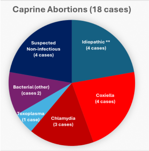
** Idiopathic includes cases in which a pathogen could not be determined either due to insufficient testing or considered not to be infectious based on histopathology and received a full diagnostic work up (Chlamydia/Coxiella/Toxoplasma PCR; bacterial culture; any additional non-routine testing) with no determined cause.
- Only 1/8 abortion cases considered “idiopathic” received a full diagnostic work up.
- 4 goat abortions were considered likely to be non-infectious based on a lack of histological lesions or a specific diagnosis of a non-infectious cause of abortion
| Ovine | Caprine | |
| Respiratory | 12 cases. Mannheimia haemolytica (1), T. pyogenes (1), aspiration pneumonia (2), “Bacterial” – not cultured (7), nasal adenocarcinoma (1). | 9 cases. “Bacterial” – not cultured (3), CAE (1), Mannheimia haemolytica (3), aspiration (1), (1). |
| CNS | 3 cases. Polioencephalomalacia (1), Listeria monocytogenes encephalitis (1), potential copper deficiency in lambs (1). | 1 case. Idiopathic (1) – considered chronic – rule out old bacterial or parasite migration tracts. Only gross and histology preformed |
| GI | 4 cases (including pathogens like ETEC, EPEC, Rotavirus, other bacteria, rumenitis, abomasitis, bloat, GI neoplasia and idiopathic – excluding parasites, C. perf, Salmonella).: 1 case of EPEC in young lambs, 1 case lamb abomasitis/rumenitis, 1 case presumed bacterial enterocolitis (no culture), 1 case oral ameloblastoma. | 3 cases. 1 attributed to bloat, 1 case of Johne’s (MAP), 1 idiopathic case suspected chronic parasite infestation. |
| Parasitology | Case numbers were decreased from previous quarters – 9 cases (11 samples) and the most frequently identified parasite by flock were 6 cases of Coccidia. There was 1 diagnosis of Cryptosporidium, 2 of Nematodirus (thread-necked), 1 of GIN, 1 Strongyloides (threads) and 1 Trichuris (whips). One case of Haemonchus was diagnosed by pathologists on postmortem. | Submissions on par with previous quarters – 24 cases (26 samples) and the most frequently identified parasites by herd including 19 cases of Coccidia, 4 of GINs, 5 of Strongyloides (threads), and 3 of Cryptosporidia. There were 2 cases of Cestodes/Moniezia (tapeworm), 1 of Trichuris (whip), 2 of Muellerius (lung worm), and 1 of Skrjabinema caprae (pinworm). |
| Clostridium perferingens | The number of isolations was similar to previous quarters. In total 11 cases isolated clostridium perfringens and only 2 were genotyped. Only 1 typed as C. perfringens type D, causing enterotoxemia (and 1 C. perfringens type A). | The number of isolations was similar to previous quarters, but with more genotyping. 25 cases isolated Clostridium perfringens and 13 were genotyped. 10 were typed as C. perfringens type D, causing enterotoxemia (1 C. perfringens type A and 2 C. perfringens type A with beta-2 toxin). |
2025 CAEV, MVV, and Johne’s Disease Report
Ovine:
MVV 133 submissions (1507 individual serum samples) submitted. There were 46 positive/suspicious cases (102 samples) and the positive case rate (one or more positive samples per case submission) was 35%.
Johne’s disease 69 submissions (303 individual samples) submitted for Johne’s disease testing. There were 2 cases (3 samples) positive by PCR and 5 cases (7 samples) positive by ELISA.
Caprine:
CAEV 154 submissions (3361 individual serum samples) submitted. There were 66 positive/suspicious cases (671 samples) and the positive case rate (one or more positive samples per case submission) was 43 %.
Johne’s disease There was a total of 175 submissions (900 individual samples) submitted. There were 18 cases (30 samples) positive by PCR and 35 cases(201samples) positive by ELISA.
Pathology Case Highlight
Signalment: Three adult Saanen goats.
Clinical history: Acute forelimb lameness progressed to 3-legged lameness resulting in euthanasia. Case was submitted for PM.
Gross lesions: Severe fibrinous and suppurative arthritis affecting the left carpus, right elbow and right fetlock; no evidence of trauma was appreciated.
Microscopic lesions: Severe suppurative and fibrinous inflammation of the synovium with occasional intralesional clusters of bacteria (not suggestive of CAEV and antigen was not detected by IHC).
Additional Testing: No mycoplasma organisms were isolated on mycoplasma culture. Bacterial culture yielded large numbers of Streptococcus dysgalactiae subsp. dysgalactiae from the left carpus and fewer from the right elbow and fetlock joints.
Diagnosis: S. dysgalactiae subsp. dysgalactiae is a relatively well-known cause of septic arthritis (“joint ill”) in young animals; however, it has also been reported to cause polyarthritis in adult goats of the Saanen breed, and this breed may be more susceptible than others.
AHL Newsletter link: https://www.uoguelph.ca/ahl/streptococcal-arthritis-adult-goat
Camelid Data Snapshot, 2024-2025
| Alpaca | Llama | Camel | |
| Parasitology | 90 samples submitted. 75 fecal egg count (53 McMaster and 22 Wisconsin), 14 fecal floatation, and 1 fecal centrifugal sedimentation. The most identified parasites were 54 Coccidia and 61 GIN. Other species also identified include Trichuris (5), Nematodirus (roundworms) (4), Capillaria (3), and Moniezia (4). 12 negative results. | 10 samples submitted. 8 McMaster fecal egg count and 2 fecal floatation. GIN was identified in 6 of the samples. Other species also identified include Strongyloides (3), Moniezia (1), Trichuris (1), Coccidia (2), Capillaria (2). 2 negative results. | 2 samples submitted for McMaster fecal egg count. GIN and Trichuris were each identified once. 1 negative result. |
Case Report: Chronic proliferative rhinitis due to Salmonella
By: Dr. Amy Gaw
A well-established producer with approximately 300 ewes in a pasture-based system, called for a ewe with marked noise on breathing. The noise happened both on inspiration and expiration, and the ewe had smooth pendulous tissue protruding from the left nostril. The ewe was euthanized and a postmortem examination was performed. Examination of the thoracic and abdominal organs revealed no gross lesions. Pink, smooth, pendulous tissue could be seen at the nares, so the head was removed and sent to AHL for further diagnostics. Enzootic nasal tumor was high on the differential diagnosis list as a potential cause.
Pathology and histology at the AHL revealed a fleshy space occupying mass in both nasal cavities. Histology revealed a hyperplastic rhinitis with numerous interepithelial bacteria. Culture revealed Salmonella as an underlying cause. Chronic proliferative rhinitis due to Salmonella infection has been documented sparingly in the literature. A second, similar case in this flock was also confirmed as Chronic proliferative rhinitis secondary to the same strain of Salmonella spp. This is a condition that should be considered by practitioners in sheep with marked inspiratory stridor. There is no known treatment or prevention for this condition currently.
Provincial Abattoir Slaughter and Condemnation Data
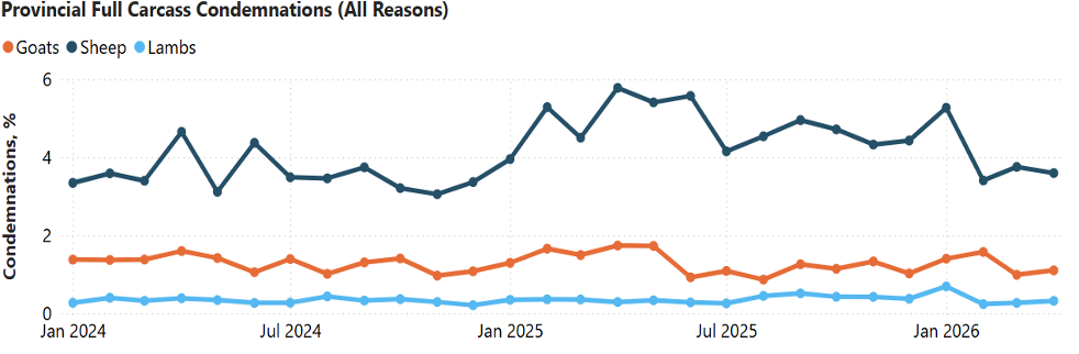
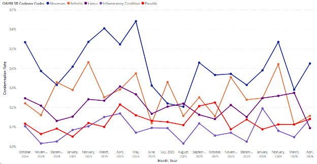
Provincial condemnation data for goat, sheep and lamb
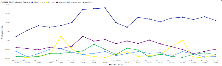
Top 5 condemnation reasons for sheep at provincial abattoirs
Top 5 condemnation reasons for lamb at provincial abattoirs
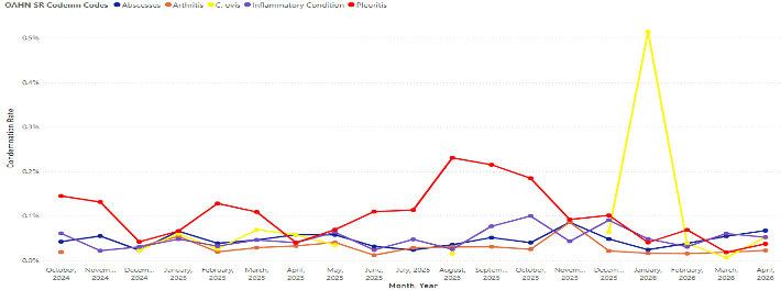
Top 5 condemnation reasons for goat at provincial abattoirs
Research Highlights
Reservoirs and proposed transmission routes of Staphylococcus aureus related to intramammary infections in dairy goats.
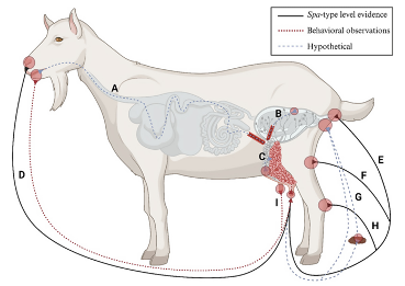
Abstract: “…In this paper, we reviewed the literature on intramammary and extramammary S. aureus reservoirs. We found 71 studies that investigated the occurrence of S. aureus intramammary infections in dairy goats showing no signs of clinical mastitis. In most of the studies (67.6%: 48 out of 71) the reported proportion of dairy goat udder halves with an S. aureus intramammary infection was below 10%. We found 9 studies that took samples of extramammary body sites of clinically healthy dairy goats. The nose had the highest proportion of positive samples for S. aureus followed by feces and rectum, and then the vulva, hock, groin, udder cleft and teat skin, in that order. The prevalence estimates reported in this review are descriptive of nature and substantial heterogeneity across studies in study design and diagnostic tools used was found. In addition, we show that the transmission routes from extramammary body sites to intramammary infections in dairy goats are not well understood…”
Pals, M. A. M., Lam, T. J. G. M., Wisselink, H. J., Smit, L. A. M., & Koop, G. (2026). Reservoirs and proposed transmission routes of Staphylococcus aureus related to intramammary infections in dairy goats: A scoping review. Journal of Dairy Science. Advance online publication. https://doi.org/10.3168/jds.2025-28203
International Disease Topics of Concern
New World screwworm resources:
USDA APHIS: Confirmed Detections of New World Screwworm | Animal and Plant Health Inspection Service
Epizootic hemorrhagic disease
Epizootic hemorrhagic disease (EHD) has been confirmed as the cause of severe neurological abnormalities in calves from multiple cattle herds in Wallowa County, Oregon. The vector-borne virus, transmitted by biting midges and more commonly affecting wild deer, has resulted in stillbirths and calves born with extensive brain necrosis, with an estimated 40–50 cases reported locally. Sheep, goats and camelids might also be susceptible but usually do not develop overt disease. There is no available vaccine or treatment, and control efforts are focused on reducing insect exposure in affected areas.
Global Expansion of Foot and Mouth Disease Serotype SAT 1
Over the past year, expansion foot-and-mouth disease virus (FMDV) serotype SAT-1 has shown unprecedented geographic, moving beyond its historic confinement to sub-Saharan Africa. Since 2025, SAT-1 has been detected across parts of Western Asia, North Africa, and southeastern Europe, with two cocirculating topotypes identified. Topotypes are genetically and geographically distinct groups within a virus serotype, defined by characteristic patterns in viral genome sequences that reflect regional evolutionary lineages. Because immunity and vaccines are serotype-specific, the spread of SAT-1 into largely naïve livestock populations has significantly heightened transboundary disease risk and surveillance concern.