Ontario Animal Health Network (OAHN)
Small Ruminant Expert Network
Report
Animal Health Laboratory (AHL) Case Summary Data April – Oct 2025
- A total of 39 ovine postmortem cases and 50 histopathology cases from both laboratory and on-farm postmortems/biopsy were submitted to the AHL and OVC-PBI from April 1 to September 30, 2025. A total of 28 of these cases had both postmortem and histology performed; comprising 72% of total postmortems submitted.
- A total of 32 caprine postmortem and 40 histopathology cases from both laboratory and on-farm postmortems/biopsy were submitted to the AHL and OVC-PBI from April 1 to September 30, 2025. A total of 26 of these cases had both postmortem and histology performed; comprising 81% of total postmortems submitted.
Abortion Case Findings
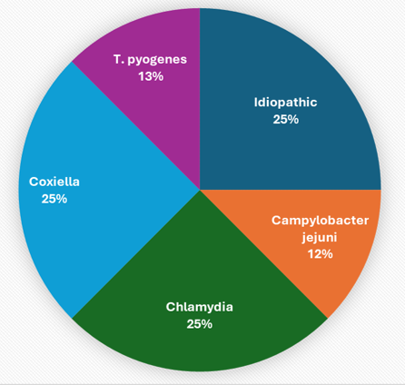
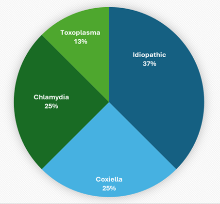
** Idiopathic includes cases considered to be non-infectious OR those in which a pathogen could not be determined either due to insufficient testing or with a full diagnostic work up including histopathology, Chlamydia/Coxiella/Toxoplasma PCR and bacterial culture or any additional non-routine testing. Only 2/5 sheep and goat cases considered “idiopathic” received a full diagnostic work up.
| Ovine | Caprine | |
| Respiratory | Pasteurella multocida (2), Mycoplasma ovipneumoniae (2), Mannheimia haemolytica (1), T. pyogenes (1), aspiration pneumonia (2), “Bacterial” – not cultured (1), idiopathic (1). The single idiopathic case was diagnosed a bronchointerstitial pneumonia of presumed viral and bacterial pathogens, but no ancillary testing was performed.
|
“Bacterial” – not cultured (6), T. pyogenes (1), E. coli (1), Mannheimia haemolytica (1), Mycoplasma ovipneumoniae (1), Bibersteinia trehalosi (1).
|
| CNS | Polioencephalomalacia (3), Listeria monocytogenes encephalitis (1).
|
Polioencephalomalacia (1), Listeria monocytogenes encephalitis (4), “bacterial” encephalitis (2). |
| GI | 5 cases. 1 case of ETEC, 1 case of rotavirus B, 3 cases of rumenitis or abomasitis, 2 cases of “bloat” with visible sarcina-like organisms seen histologically. | 3 were attributed to bloat, 1 to Johne’s (MAP) and 1 idiopathic. The idiopathic case was a fibrinonecrotizing ileitis/typhlitis, but no histology or ancillary testing performed
|
| Parasitology | 23 cases (34 samples). As usual, the most frequently identified parasites by flock/case were 24 cases of Coccidia and 10 of GIN eggs. There were 4 diagnoses of Cestodes (tapeworms), 1 Cryptosporidium, 2 of Nematodirus (roundworms), 6 Strongyloides (threads) and 3 Trichuris (whips). Six cases of Haemonchosis were diagnosed by pathologists on postmortem.
|
28 cases (42 samples) and the most frequently identified parasites by flock including Coccidia (21 cases), GINs (14), 4 of Strongyloides (threads), and Cryptosporidia (4). There was 1 case of Cestode/Moniezia (tapeworm), 1 of Trichuris (whip), 3 Muellerius (lung worm), and 2 of Skrjabinema caprae (pinworm). Giardia cysts were seen on fecal float in a single case. One case of Dicrocoelium (lancet fluke) was diagnosed histologically. One case of haemonchosis was diagnosed by pathologists.
|
| Clostridium perferingens | The number of isolations were similar to previous quarters. In total 14 cases isolated C. perfringens and only 5 were genotyped. Only 2 typed as C. perfringens type D causing enterotoxemia. | The number of isolations were similar to previous quarters. 19 cases isolated C. perfringens and only 2 were genotyped. One was typed as C. perfringens type D causing enterotoxemia (1 C. perfringens type A with Beta-2 toxin).
|
Pathology Case Highlight: Ovine White Liver
Signalment: Yearling ewe (submitted for postmortem examination).
Clinical history: Several weeks anorexia, depression, and weight loss. Several animals had died sporadically in the past year with similar symptoms.
Gross lesions: Thin and watery blood (supporting anemia), mucus membranes off-white (no evidence of jaundice), severe effusion in the abdomen, thorax, and pericardium, and liver diffusely pale and very firm.
Microscopic lesions: Severe bridging fibrosis of the liver, abundant hepatocellular lipid along with ceroid pigment and scattered neutrophils. Hypertrophy of cerebral astrocytes (neuronal support cells) in the brain (suggestive of hepatic encephalopathy).
Diagnosis: With additional testing, liver cobalt levels too low to be detected.
Ovine white liver disease is a metabolic disorder in sheep caused by a deficiency in Vitamin B12, which is synthesized by the gut microbes using dietary cobalt. Low levels associated with sandy or high pH soil, or intensive cropping causing leaching of the soil, or lack of cobalt enriched blue salt lick. Studies of lambs with experimentally induced white liver show reduced activity of vitamin B12-dependent enzymes within hepatocytes à hinders the oxidation of fatty acids and is the probable cause of lipid accumulation and secondary hepatocellular injury. Symptoms/lesions of hepatic encephalopathy are occasionally reported. Liver damage causes an increased levels of circulating ammonia leading to brain injury.
AHL Newsletter link: https://www.uoguelph.ca/ahl/ovine-white-liver-disease-yearling-ewe
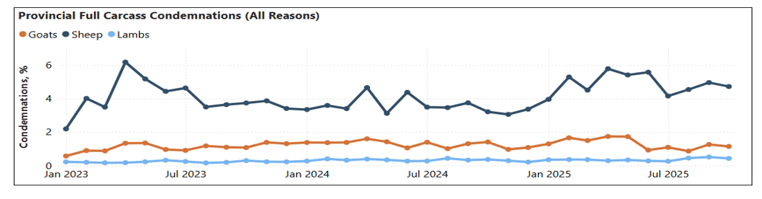
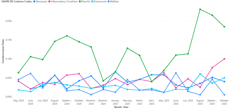
Provincial Abattoir Slaughter and Condemnation Data
Provincial condemnation data for goat, sheep and lamb, Jan 2023 to Oct 2025.
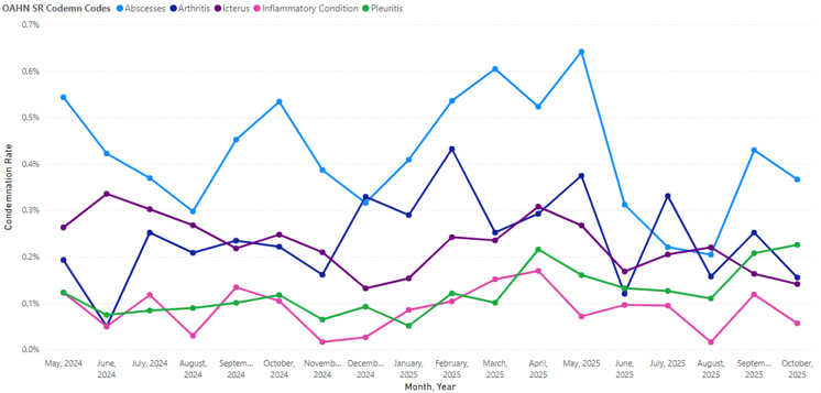
Top 5 condemnation reasons for sheep at provincial abattoirs May 2024-Oct 2025.
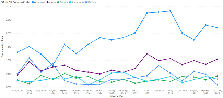
Top 5 condemnation reasons for lamb at provincial abattoirs May 2024-Oct 2025.
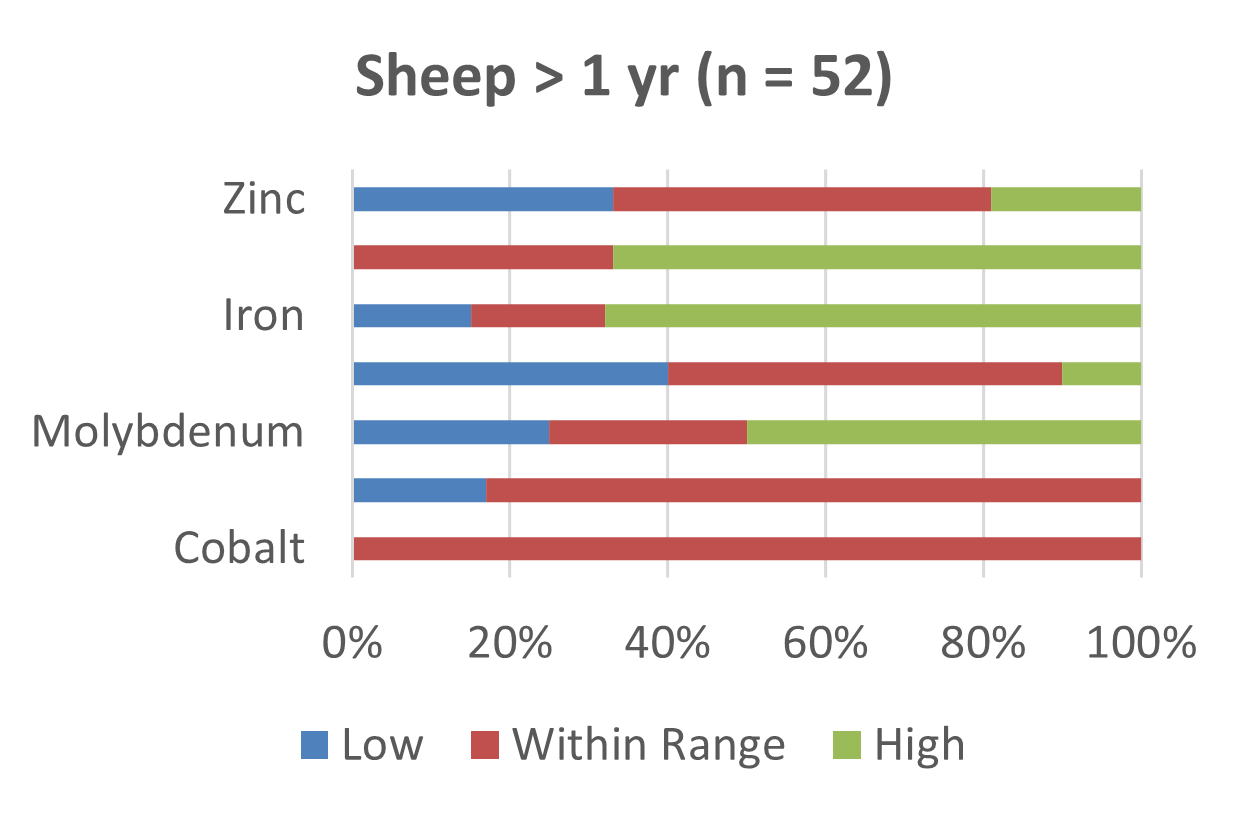
Small Ruminant Trace Mineral Analysis 2020-2025 by AHL
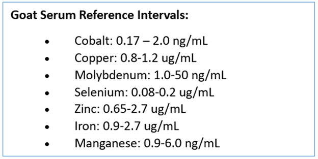
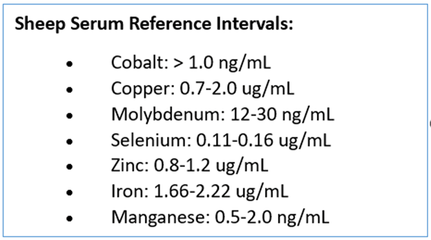
This quarter, the small ruminant OAHN network reviewed trace mineral testing data in serum samples from the Animal Health Lab. Common deficiencies in both goat kids and adults included zinc, copper and selenium (Figures 1 & 2). Both age groups in sheep showed a broader range of mineral deficiencies than goats including iron, copper, zinc and selenium (Figures 3 & 4). In addition, molybdenum was above the reference interval in 20% of lambs and 30% of adult sheep. No goats tested above the reference interval for molybdenum. This data may include animals with and without mineral supplementation as history was not provided in most submissions.

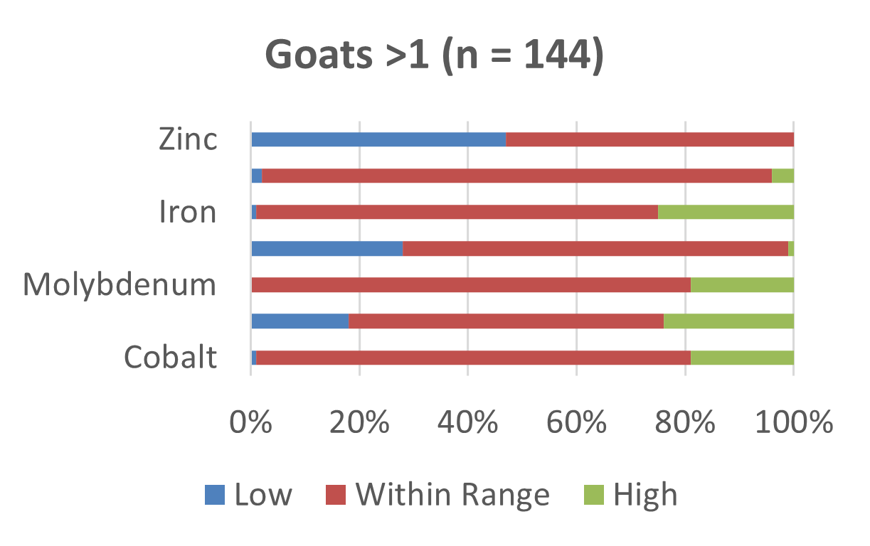
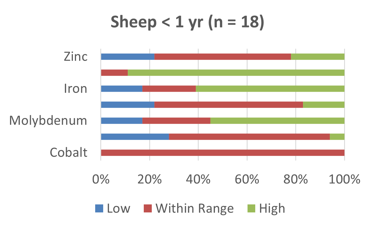
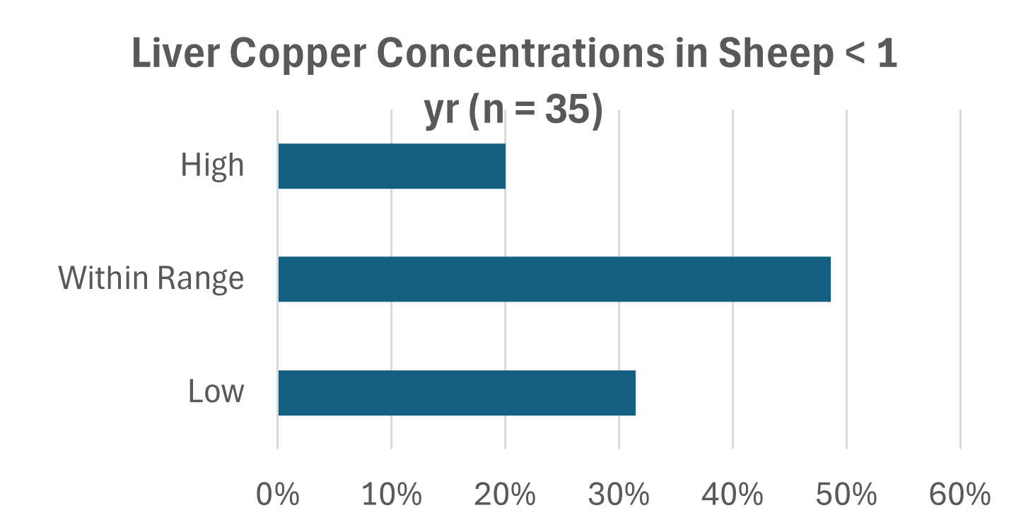
Based on feedback from field veterinarian reports of copper deficiency in lambs data from copper testing on liver samples was analysed (Figures 5).
Case Report: Malignant catarrhal fever in Icelandic sheep
By: Francesca Ruberto, OVC class of 2026
This case report presents a rare occurrence of clinical manifestation of MCF in Icelandic lambs and one ram.
Seven Icelandic lambs ranging from two to seven weeks of age, and one Icelandic ram of one year of age, on a mixed-breed sheep farm, developed overlapping multisystemic clinical signs. The main clinical signs included pyrexia, lethargy, mucopurulent ocular/nasal/urogenital discharge, diarrhea, coughing, dyspnea, mucocutaneous lesions and painful urination and/or defecation. Sheep of the Icelandic breed were the only breed affected. Clinical progression ranged from mild respiratory disease to severe systemic signs. Two of the lambs died, four were humanely euthanized, and one lamb and the adult ram survived with aggressive supportive care. The main therapeutic interventions used were meloxicam, florfenicol and penicillin.
The deceased lamb was submitted to AHL. The post-mortem examination reported death secondary to shock related to intussusception. The histopathology results indicated enterocolitis, glandular dropout, lympholysis of Peyer’s patches, focal necrosis of a lymph node, marked ulcerative esophagitis, marked neutrophilic rumen reticulo-omasitis, lympholysis of the thymus and spleen, mild multifocal cardiac and skeletal lymphocytic myositis, mild lymphocytic tracheitis, and mild pulmonary edema. An MCF-associated OvHV-2 PCR of the splenic tissue was positive with a low Cycle threshold (Ct) of 27.66, leading to a diagnosis of MCF.
The primary MCF pathology includes lymphoproliferative inflammatory infiltrates, systemic vasculitis and epithelial necrosis1,3. Unfortunately, confirmation of naturally occurring MCF in sheep is a problem as detection of OvHV-2 specific antibodies or DNA are often positive in sheep despite lack of clinical signs1,4. Previous studies have concluded that high copy numbers of viral DNA in tissues that are associated with lesions characteristic to MCF can be used to confirm the diagnosis of MCF in sheep1. In this case study, the lamb exhibited a low OvHV-2 Ct in the spleen. The low Ct value and histopathology results paired with the SA-MCF related clinical signs strongly support a diagnosis of MCF in this case.
References
1. Phillips H, Hughes KJ, Little PB, Hayes MA. High copy number of ovine gammaherpesvirus 2 DNA associated with malignant catarrhal fever–like syndrome in a lamb. J Vet Diagn Invest 2018;30(4):623–627.
2. Rahim M, Li H, Taus NS, Lewis GS, Kim O, Traul DL, Crawford TB. The effect of age on the excretion of ovine herpesvirus-2 (OvHV-2), the causative agent of malignant catarrhal fever (MCF), in naturally infected sheep. Vet Microbiol 2010;142(3-4):344–349.
3. Li H, Taus NS, Lewis GS, Kim O, Traul DL, Crawford TB. Shedding of ovine herpesvirus 2 in sheep nasal secretions: the predominant mode for transmission. J Clin Microbiol 2004;42(12):5558–5564.
4. Bremer CW. The prevalence of ovine herpesvirus-2 in 4 sheep breeds from different regions in South Africa. J S Afr Vet Assoc 2010;81(2):93–96.
International Disease Topics of Concern
The Asian longhorned tick (Haemaphysalis longicornis) is an invasive species that has rapidly expanded its range since its first detection in the United States in 2017. Native to East Asia, this tick has now been found in multiple U.S. states, including New York, Pennsylvania, Ohio and Michigan — regions not far from the Ontario border. Most recently, it was detected in Maine and Kansas. Although it has not yet been detected in Canada, its potential to establish in Ontario is a growing concern among public health and veterinary experts.
For more information, see OAHN’s new factsheet: Asian Longhorned Tick- An encroaching threat
Research Highlight
“We inoculated two groups of lactating goats via intramammary and respiratory routes with Cow-H5N1 (genotype B3.13) or avian-H5N1 (genotype B1.2) virus. Both groups developed severe clinical mastitis and shed viruses in milk, resulting in transmission to suckling kids. Viral RNA was detected in nasal and oral swabs and various tissues, and virus-neutralizing antibodies were present in serum, milk, and bronchoalveolar lavage fluid. In vitro, both viruses replicated efficiently in goat respiratory and mammary epithelial cells. Mammary tissue expresses both α2,3- and α2,6-linked sialic acid receptors. These findings demonstrate that goats are highly susceptible to H5N1 infection, with mammary tropism facilitating transmission to offspring, and underscore the need for increased surveillance in ruminant livestock.”
Alkie, Tamiru N et al. “Dairy cow- and avian-origin clade 2.3.4.4b H5N1 induce severe mastitis in lactating goats and transmission to suckling goats.” Cell reports vol. 44,10 (2025): 116346. doi:10.1016 https://doi.org/10.1016/j.celrep.2025.116346